


LEARNING OBJECTIVES
After studying this topic, students will be able to:
- Explain neuronal inhibition and IPSP.
- Differentiate presynaptic and postsynaptic inhibition.
- Explain spatial summation, temporal summation, and facilitation.
- Describe the special role of dendrites in neuronal excitation and inhibition.
- Explain electrotonic and decremental conduction in dendrites.
INTRODUCTION
Neurons continuously receive both excitatory and inhibitory signals. Excitatory signals move the membrane potential toward the firing threshold, whereas inhibitory signals make the membrane potential more negative and reduce the chance of firing. The final response of a neuron depends on how these signals add together, oppose each other, and spread through the dendrites and soma. Spatial summation, temporal summation, facilitation, electrotonic conduction, and dendritic inhibition all help determine whether the neuron will finally generate an action potential.
SPATIAL SUMMATION IN NEURONS—THRESHOLD FOR FIRING
- Excitation of only one presynaptic terminal almost never excites the neuron.
- A single terminal usually produces an EPSP of only about 0.5–1 mV.
- However, about 10–20 mV is normally needed to reach the threshold for excitation.
- Therefore, one presynaptic terminal usually does not produce enough depolarization to fire the neuron.
- Usually, many presynaptic terminals are stimulated at the same time.
- These terminals may be spread over different areas of the neuron, but their effects can still add together.
- As more excitatory synapses discharge at the same time, the total potential inside the soma becomes more positive.
- Each simultaneously active excitatory synapse can increase the intrasomal potential by about 0.5–1 mV.
- When the total EPSP becomes large enough, the neuron reaches the firing threshold.
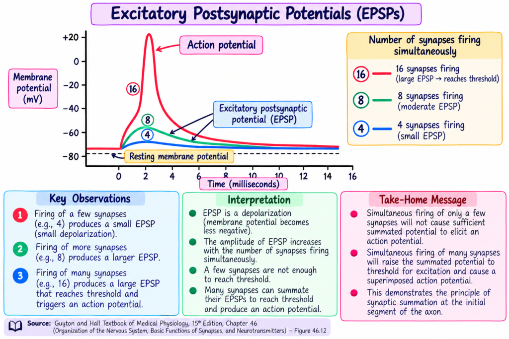
- At threshold, an action potential develops spontaneously in the initial segment of the axon, as shown in Fig. 46.12.
- In Fig. 46.12:
- Stimulation of 4 synapses produces the lowest postsynaptic potential.
- Stimulation of 8 synapses produces a higher postsynaptic potential.
- Stimulation of 16 synapses produces an even higher EPSP.
- With 16 synapses, the firing threshold is reached and an action potential is generated in the axon.
- The addition of simultaneous postsynaptic potentials from multiple terminals located over different areas of the neuronal membrane is called spatial summation.
Shortest -Story Version
Imagine a neuron receiving many tiny excitatory signals at the same time. One synapse gives only a very small EPSP, so it usually cannot make the neuron fire. But when many synapses are active together, their small EPSPs add up. As the total EPSP becomes larger, it reaches the firing threshold, and then an action potential starts in the initial segment of the axon. This adding of signals from different places on the neuron is called spatial summation.
KEY CONCEPT
One synapse → very small EPSP → usually no firing
Many synapses active together → EPSPs add together → threshold reached → action potential generated
Easiest Example
Think of each excitatory synapse as adding a small amount:
0.5–1 mV + 0.5–1 mV + 0.5–1 mV + more inputs → total EPSP becomes larger → threshold is reached → neuron fires
So, spatial summation means many excitatory inputs acting at the same time add together to make the neuron fire.
“TEMPORAL SUMMATION” CAUSED BY SUCCESSIVE DISCHARGES OF A PRESYNAPTIC TERMINAL
- Each time a presynaptic terminal fires, it releases a transmitter substance.
- This transmitter opens membrane channels for only about 1–2 milliseconds.
- However, the changed postsynaptic potential can remain for up to 15 milliseconds after the channels have closed.
- Therefore, if the same presynaptic terminal fires again quickly, the second postsynaptic potential can add to the first one.
- If stimulation becomes more rapid, the postsynaptic potential becomes greater.
- So, repeated rapid discharges from the same presynaptic terminal can add together.
- This adding of successive postsynaptic potentials is called temporal summation.
SIMULTANEOUS SUMMATION OF INHIBITORY AND EXCITATORY POSTSYNAPTIC POTENTIALS
- An IPSP tends to make the membrane potential more negative.
- At the same time, an EPSP tends to make the membrane potential more positive.
- When both occur together, they can partially or completely cancel each other.
- Therefore, if a neuron is being excited by an EPSP, an inhibitory signal can reduce the postsynaptic potential.
- If the postsynaptic potential falls below the threshold for excitation, the activity of the neuron can be turned off.
KEY CONCEPT
Temporal summation:
Same presynaptic terminal fires rapidly again and again → postsynaptic potentials overlap → they add together → larger postsynaptic potential
Simultaneous excitation and inhibition:
EPSP increases membrane potential + IPSP decreases membrane potential → both oppose each other → neuron may fail to reach threshold
Easiest Example
If one terminal sends signals rapidly one after another, each new signal arrives before the previous postsynaptic effect has fully disappeared. The effects therefore add together. This is temporal summation.
If an EPSP and IPSP occur at the same time, one pushes the membrane potential upward while the other pulls it downward, so they can reduce or cancel each other’s effect.
FACILITATION OF NEURONS
- Sometimes the summated postsynaptic potential is excitatory, but it is still not strong enough to reach the firing threshold.
- In this condition, the neuron is said to be facilitated.
- This means the membrane potential has moved closer to the threshold for firing than normal.
- However, the neuron has not yet reached the firing level.
- Because the neuron is already close to threshold, another excitatory signal coming from a different source can excite the neuron very easily.
- Diffuse signals in the nervous system often facilitate large groups of neurons.
- This allows these neurons to respond quickly and easily to signals arriving from other sources.
Shortest – Story Version
A neuron may receive enough excitatory signals to move close to threshold, but not enough to fire. At this stage, the neuron is facilitated. Because it is already near threshold, one more excitatory signal from another source can make it fire very easily.
KEY CONCEPT
Excitatory inputs add together → membrane potential moves near threshold → neuron does not fire yet → neuron becomes facilitated → another small excitatory signal can easily trigger firing
Easiest Example
Think of the neuron as being almost ready to fire. It has not reached threshold yet, but it is very close. If one more excitatory signal arrives, the neuron can fire easily. This condition is called facilitation.
SPECIAL FUNCTIONS OF DENDRITES FOR EXCITING NEURONS
- The dendrites of anterior motor neurons often extend about 500–1000 micrometers in all directions from the neuronal soma.
- Because dendrites spread over a large area, they can receive signals from a large spatial field around the motor neuron.
- This gives a large opportunity for summation of signals coming from many separate presynaptic nerve fibers.
- About 80%–95% of presynaptic terminals of the anterior motor neuron end on the dendrites.
- In contrast, only about 5%–20% of presynaptic terminals end on the neuronal soma.
- Therefore, a large share of neuronal excitation is produced by signals transmitted through the dendrites.
KEY CONCEPT
Wide dendritic spread → receives signals from a large area → many presynaptic inputs can add together → strong contribution to neuronal excitation
Easiest Example
Think of dendrites like many wide branches of a tree. Because they spread far around the neuron, they can collect signals from many different nerve fibers. Since most presynaptic terminals end on dendrites, dendrites provide a major part of the neuron’s excitation.
MOST DENDRITES CANNOT TRANSMIT ACTION POTENTIALS BUT THEY CAN TRANSMIT SIGNALS WITHIN THE SAME NEURON BY ELECTROTONIC CONDUCTION
- Most dendrites cannot transmit action potentials.
- This is because their membranes have relatively few voltage-gated sodium channels.
- Their threshold for excitation is also too high for action potentials to occur.
- However, dendrites can still transmit electrotonic current toward the soma.
- Electrotonic conduction means the electrical current spreads directly through the fluids of the dendrites by ion conduction.
- During this type of conduction, no action potential is generated.
- This electrotonic current can produce either stimulation or inhibition of the neuron.
KEY CONCEPT
Few voltage-gated Na⁺ channels + high excitation threshold → dendrites usually cannot produce action potentials → electrical current still spreads through dendrites → reaches the soma by electrotonic conduction
Easiest Example
Think of the dendrite as carrying a local electrical signal toward the cell body. The signal can travel through the dendrite, but the dendrite usually does not generate an action potential of its own.
DECREMENT OF ELECTROTONIC CONDUCTION IN THE DENDRITES CAUSES GREATER EXCITATORY (OR INHIBITORY) EFFECT BY SYNAPSES LOCATED NEAR THE SOMA
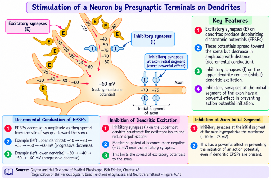
- Fig. 46.13 shows multiple excitatory and inhibitory synapses acting on the dendrites of a neuron.
- On the two dendrites on the left, excitatory effects are present near the tip ends.
- At these tip ends, the EPSPs are high, meaning the membrane potentials are less negative.
- However, a large part of the EPSP is lost before it reaches the soma.
- This happens because dendrites are long and their membranes are thin.
- Their membranes are also partly permeable to potassium and chloride ions.
- Therefore, dendrites are relatively leaky to electrical current.
- As the excitatory potential travels toward the soma, part of it is lost by leakage through the membrane.
- This gradual decrease in membrane potential during electrotonic spread toward the soma is called decremental conduction.
- The farther an excitatory synapse is from the soma, the greater the decrement.
- Therefore, less excitatory signal reaches the soma from synapses located far away.
- Synapses located near the soma have a much greater effect on neuronal excitation or inhibition.
- Synapses located far from the soma have a smaller effect.
KEY CONCEPT
Signal starts in dendrite → some electrical current leaks out → signal becomes weaker as it travels → farther synapse = more signal loss → near-soma synapse = stronger effect
Easiest Example
A synapse close to the soma sends a signal over a short distance, so less of the signal is lost. A synapse far from the soma sends the signal over a longer dendrite, so more of the signal leaks away before reaching the soma. Therefore, near synapses have a stronger effect than distant synapses.
SUMMATION OF EXCITATION AND INHIBITION IN DENDRITES
- The uppermost dendrite in Fig. 46.13 is stimulated by both excitatory and inhibitory synapses.
- At the tip of the dendrite, there is a strong EPSP.
- Nearer the soma, two inhibitory synapses act on the same dendrite.
- These inhibitory synapses produce a hyperpolarizing voltage.
- This hyperpolarizing effect can completely cancel the excitatory effect.
- It can also send a small amount of inhibition toward the soma by electrotonic conduction.
- Therefore, dendrites can summate EPSPs and IPSPs, just like the soma.
- Fig. 46.13 also shows several inhibitory synapses directly on the axon hillock and initial axon segment.
- Inhibition at this location is especially powerful.
- This is because it directly increases the threshold for excitation at the point where the action potential is normally generated.
KEY CONCEPT
EPSP + IPSP on the same dendrite → their effects add together → inhibition may cancel excitation → final effect travels toward the soma
Inhibitory synapse near axon hillock/initial segment → stronger inhibition → threshold increases where action potential normally begins
Easiest Example
A dendrite may receive a strong excitatory signal, but if inhibitory synapses act closer to the soma, they can cancel that excitation. If inhibition occurs near the axon hillock, it is even more powerful because it acts directly where the neuron normally starts the action potential.
EXCITATION STATE OF THE NEURON AND RATE OF FIRING
- At any moment, a neuron receives both excitatory and inhibitory effects.
- If excitation is greater than inhibition, the neuron is in an excitatory state.
- If inhibition is greater than excitation, the neuron is in an inhibitory state.
- When the excitatory state rises above the threshold, the neuron starts firing.
- The neuron continues to fire repeatedly as long as the excitatory state remains above the threshold.
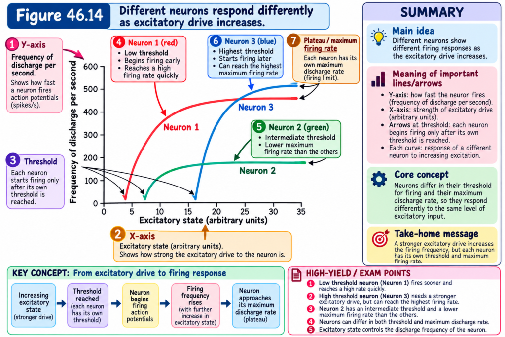
- Fig. 46.14 shows the responses of three types of neurons at different levels of excitatory state.
- Neuron 1 has a low threshold for excitation.
- Neuron 3 has a high threshold for excitation.
- However, the neurons also differ in their maximum frequency of discharge.
- Neuron 2 has the lowest maximum firing frequency.
- Neuron 3 has the highest maximum firing frequency.
- Some neurons in the central nervous system fire continuously because their normal excitatory state is already above the threshold level.
- Their firing frequency can increase further when their excitatory state increases.
- Their firing frequency can decrease when an inhibitory state is added.
- If inhibition becomes strong enough, the neuron may stop firing completely.
- Therefore, different neurons have:
- Different responses
- Different thresholds for excitation
- Different maximum frequencies of discharge
- These different response characteristics allow neurons to perform the widely varying functions of the nervous system.
KEY CONCEPT
More excitation than inhibition → Excitatory state → Threshold is crossed → Neuron fires repeatedly
More inhibition than excitation → Inhibitory state → Firing decreases or may stop
Easiest Examples
- Neuron 1: Needs a lower level of excitation to reach its threshold.
- Neuron 3: Needs a higher level of excitation to reach its threshold.
- Neuron 2: Has the lowest maximum firing frequency.
- Neuron 3: Has the highest maximum firing frequency.
- A neuron that is already above threshold can fire continuously, but added inhibition can slow or stop its firing.
PHYSIOLOGICAL IMPORTANCE
- Inhibitory synapses control neuronal firing by making the membrane potential more negative.
- Chloride influx and potassium efflux produce hyperpolarization, which reduces neuronal excitability.
- Presynaptic inhibition reduces transmission before the signal reaches the postsynaptic neuron.
- In sensory pathways, presynaptic inhibition helps reduce sideways spread and mixing of signals.
- EPSPs and IPSPs can oppose each other, allowing the nervous system to control whether a neuron reaches threshold.
- Spatial summation allows many weak inputs from different locations to combine and produce neuronal excitation.
- Temporal summation allows repeated rapid signals from one presynaptic terminal to add together.
- Facilitation prepares neurons to respond more easily because their membrane potential is already closer to threshold.
- Dendrites provide a large surface for receiving signals from many presynaptic fibers.
- Because 80%–95% of presynaptic terminals of anterior motor neurons end on dendrites, dendrites provide a large share of neuronal excitation.
- Electrotonic conduction allows dendrites to transmit electrical signals toward the soma without generating action potentials.
- Decremental conduction makes distant synaptic signals weaker before they reach the soma.
- Therefore, synapses located near the soma have a stronger excitatory or inhibitory effect than those located farther away.
- Dendrites can summate EPSPs and IPSPs, helping determine the final effect reaching the soma.
- Inhibitory synapses at the axon hillock and initial axon segment are especially powerful because they increase the threshold at the site where the action potential is normally generated.
HIGH-YIELD POINTS
- Resting neuronal membrane potential: about −65 mV.
- Chloride Nernst potential: about −70 mV.
- Opening Cl⁻ channels causes chloride influx and makes the inside more negative.
- Opening K⁺ channels causes potassium efflux and also makes the inside more negative.
- Increased intracellular negativity is called hyperpolarization.
- A more negative postsynaptic potential is called an IPSP.
- In Fig. 46.11C, membrane potential changes from −65 mV to −70 mV, producing an IPSP of −5 mV.
- Presynaptic inhibition commonly involves GABA, which opens anion channels and allows chloride ions to enter the presynaptic terminal.
- EPSP and IPSP channel effects usually last only 1–2 ms, but the postsynaptic potential may last about 15 ms.
- A single excitatory terminal usually produces only about 0.5–1 mV EPSP.
- About 10–20 mV is normally required to reach firing threshold.
- Spatial summation: signals from many presynaptic terminals at different locations add together.
Reference: Guyton and Hall Textbook of Medical Physiology, 15th Edition, Chapter 46 — Organization of the Nervous System, Basic Functions of Synapses, and Neurotransmitters.
