


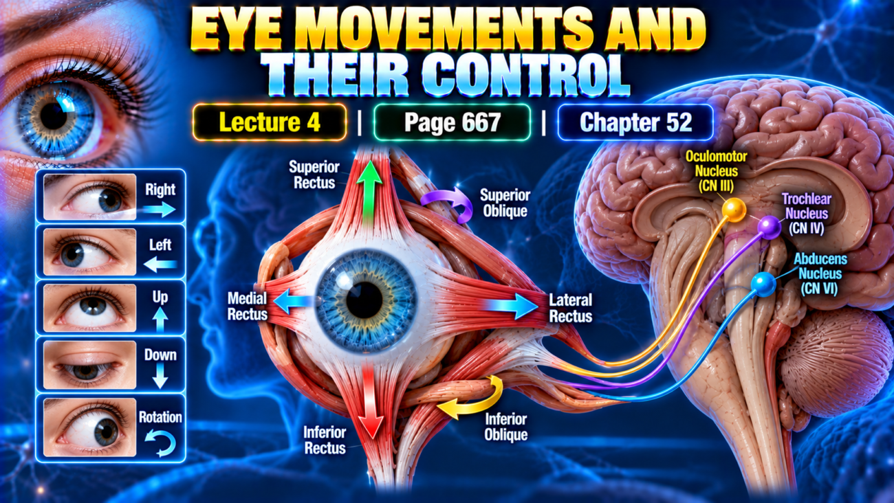
Learning Objectives
After completing this lecture, students will be able to:
- Explain the muscular and neural control of eye movements.
- Differentiate voluntary fixation, involuntary fixation, saccades, and pursuit movements.
- Explain fusion and stereopsis.
- Describe the role of the superior colliculi in orienting movements.
- Explain strabismus and suppression of vision from a repressed eye.
Introduction
Full use of vision requires a cerebral control system that directs the eyes toward the object to be viewed.
Normal eye movements depend on coordinated activity of:
- Extraocular muscles.
- Brain stem oculomotor nuclei.
- Visual cortical areas.
- Superior colliculi.
- Vestibular nuclei.
These systems allow the eyes to:
- Move toward a visual target.
- Maintain fixation.
- Follow moving objects.
- Shift between fixation points.
- Fuse the images from both eyes.
- Help judge the distance of objects.
Main Topic / Core Concept
Muscular Control of Eye Movements
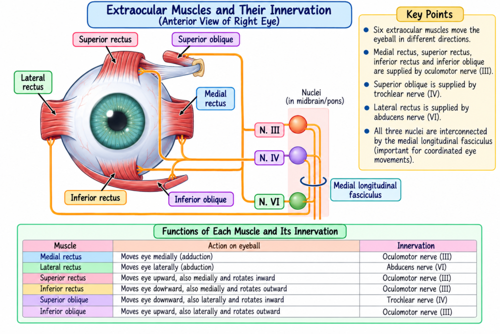
Eye movements are controlled by three pairs of muscles, shown in Fig. 52.7.
| Muscle Pair | Main Function |
|---|---|
| Medial and lateral recti | Move the eyes from side to side |
| Superior and inferior recti | Move the eyes upward or downward |
| Superior and inferior obliques | Mainly rotate the eyeballs to keep the visual fields upright |
Medial and Lateral Recti
- Control side-to-side movements of the eyes.
Superior and Inferior Recti
- Move the eyes:
- Upward.
- Downward.
Superior and Inferior Obliques
- Mainly rotate the eyeballs.
- Help keep the visual fields in the upright position.
Neural Pathways for Control of Eye Movements
The brain stem contains nuclei for the:
- Third cranial nerve.
- Fourth cranial nerve.
- Sixth cranial nerve.
These nuclei connect through peripheral nerves to the ocular muscles.
The nuclei also communicate through the:
Medial longitudinal fasciculus
Medial longitudinal fasciculus: A nerve tract connecting the brain stem nuclei involved in eye movements.
Reciprocal Innervation
Reciprocal innervation: Coordinated activity in which one muscle of an opposing pair relaxes while the other contracts.
Flow
One ocular muscle contracts
→ opposing muscle relaxes
→ coordinated eye movement
Cortical Control of Eye Movements
Cortical control is illustrated in Fig. 52.8.
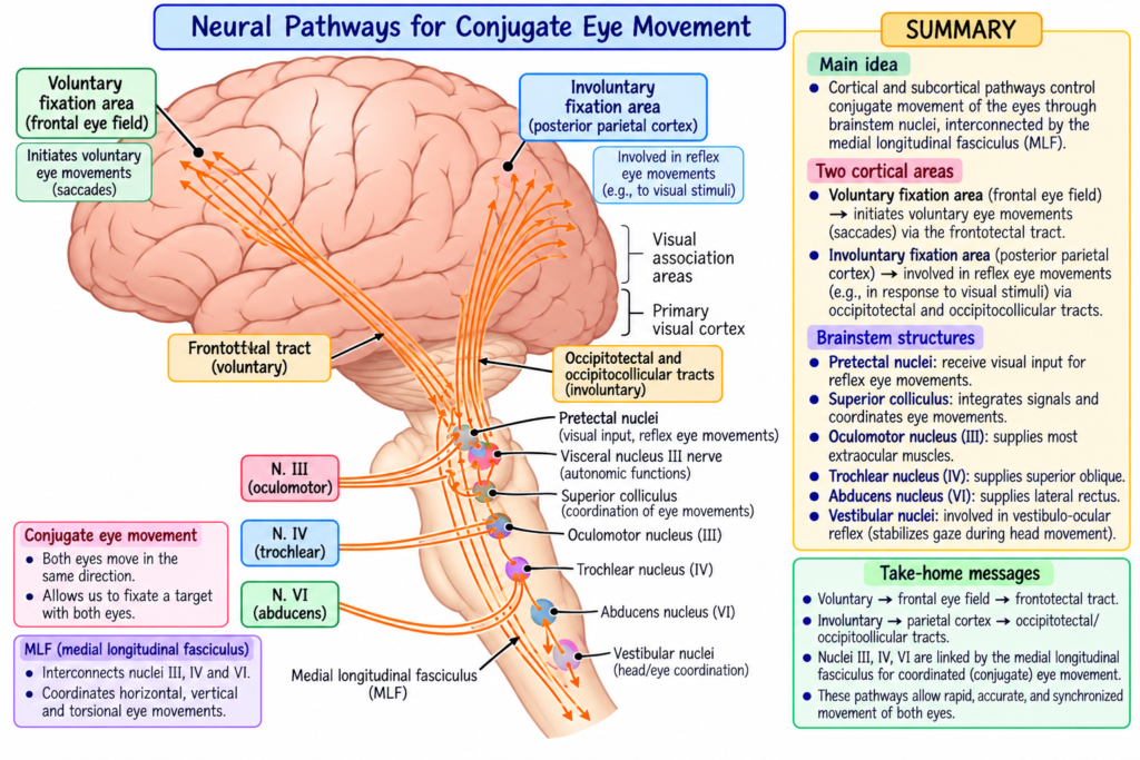
Visual areas of occipital cortex
→ occipitotectal and occipitocollicular tracts
→ pretectal area + superior colliculus
→ brain stem oculomotor nuclei
→ ocular muscles
Strong signals also arise from the vestibular nuclei.
Vestibular nuclei: Brain stem equilibrium control centers that send signals into the oculomotor system.
Vestibular Pathway
Vestibular nuclei
→ medial longitudinal fasciculus
→ oculomotor system
Fixation Movements of the Eyes
Fixation: Directing the eyes toward and maintaining them on a particular part of the visual field.
Fixation depends on two neuronal mechanisms:
- Voluntary fixation.
- Involuntary fixation.
Voluntary Fixation
Voluntary fixation mechanism: Allows a person to move the eyes voluntarily to find an object on which to fix vision.
It is controlled by:
- Bilateral cortical fields.
- Premotor cortical regions of the frontal lobes.
Function
Present fixation point
→ voluntary eye movement
→ new object located
→ new fixation established
If these frontal areas are damaged bilaterally:
- It becomes difficult to unlock the eyes from one fixation point.
- Moving the eyes to another fixation point becomes difficult.
- Blinking or briefly covering the eyes may allow the eyes to move again.
Involuntary Fixation
Involuntary fixation mechanism: Keeps the eyes firmly directed toward an object after it has been found.
It is controlled mainly by:
- Secondary visual areas of the occipital cortex.
- These areas lie mainly anterior to the primary visual cortex.
If these areas are destroyed bilaterally: Voluntary vs Involuntary Fixation
| Feature | Voluntary Fixation | Involuntary Fixation |
|---|---|---|
| Main function | Finds a new fixation point | Holds the eyes on the selected point |
| Main cortical region | Premotor frontal cortex | Secondary occipital visual areas |
| Basic action | Unlocks fixation | Locks fixation |
| Bilateral dysfunction | Difficulty shifting fixation | Difficulty maintaining fixation |
Mechanism / Pathway / Step-by-Step Explanation
Involuntary Locking Fixation
Involuntary fixation works through a negative feedback mechanism.
Negative feedback mechanism: A mechanism that prevents the object of attention from leaving the foveal region.
Flow
Image begins moving away from foveal center
→ corrective eye movement
→ image moves back toward the center of the fovea
Continuous Small Eye Movements
The eyes normally show three continuous but almost imperceptible movements:
- Tremor.
- Slow drift.
- Sudden flicking movements.
Tremor
Tremor: Continuous small eye movement occurring at approximately 30–80 cycles/sec.
It results from:
- Successive contractions of motor units in the ocular muscles.
Slow Drift
Slow drift: Slow movement of the eyeballs in one direction or another.
Flicking Movements
Flicking movements: Sudden corrective movements controlled by the involuntary fixation mechanism.
When a spot approaches the edge of the fovea:
Spot drifts toward foveal edge
→ sudden reflex reaction
→ flicking movement
→ spot moves back toward the foveal center
The drifting and flicking movements are shown in Fig. 52.9.
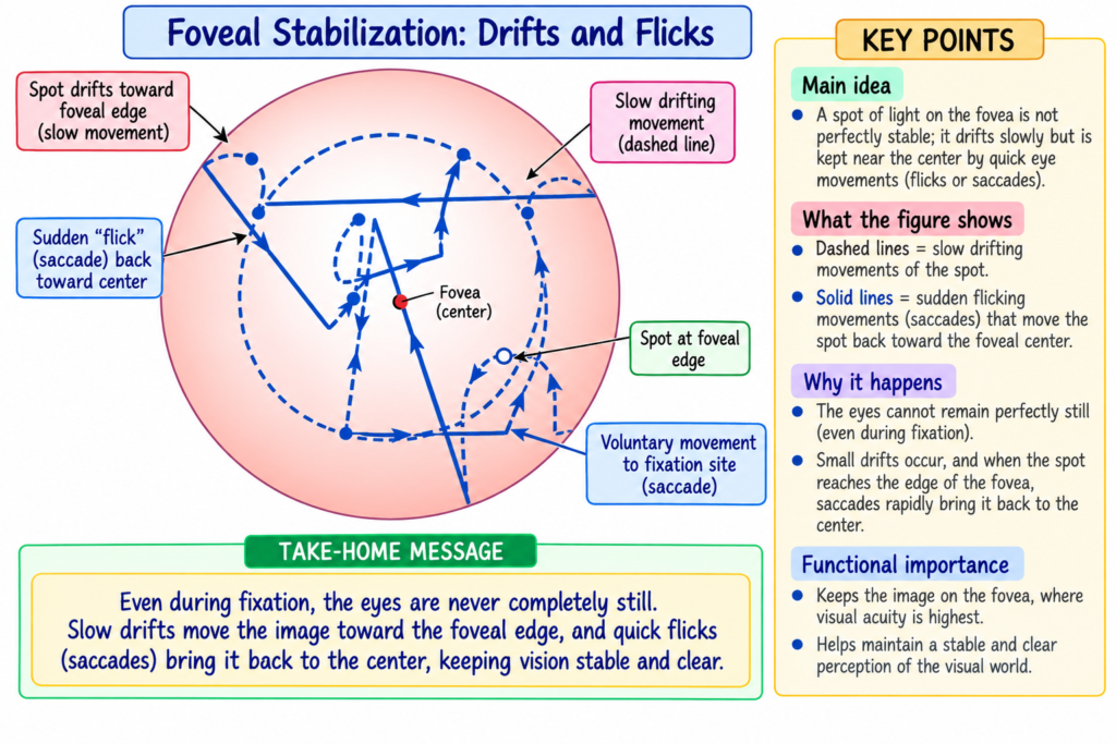
- Dashed lines → slow drifting.
- Solid lines → corrective flicking movements.
The involuntary fixation mechanism is mostly lost when the superior colliculi are destroyed.
Saccadic Movements of the Eyes
Saccade: A rapid eye movement that shifts fixation from one visual point to another.
When a visual scene continuously moves before the eyes:
- The eyes fix on one visual highlight.
- Then jump rapidly to another.
- This occurs approximately 2–3 times per second.
These rapid jumps are called:
- Saccades
The movements described in this situation are called:
- Opticokinetic movements
Time Distribution During Saccades
Approximately:
- No more than 10% of the time is spent moving the eyes.
- About 90% is spent at fixation points.
Flow
Fixation point A
→ rapid saccade
→ fixation point B
→ rapid saccade
→ fixation point C
During saccades:
- The brain suppresses the visual image.
- Therefore, the person does not consciously see the rapid movements between fixation points.
Saccadic Movements During Reading
During reading:
- Several saccades occur along each line.
- The scene is stationary.
- The eyes move across it by successive saccades.
- This helps extract important visual information.
Flow
Part of line
→ fixation
→ saccade
→ next part
→ fixation
Saccades While Viewing a Painting
When viewing a painting, saccades may move:
- Upward.
- Sideways.
- Downward.
- At different angles.
The eyes move from one visual highlight to another.
Pursuit Movement
Pursuit movement: Eye movement that allows the eyes to remain fixed on a moving object.
A cortical mechanism:
- Detects the direction or course of the moving object.
- Develops a similar movement pattern for the eyes.
Development of Pursuit
Moving object
→ eyes initially may not follow accurately
→ saccadic jumps begin to approximate its movement
→ movements become progressively smoother
→ eyes eventually follow the object almost exactly
This represents a high degree of automatic subconscious control.
Saccades vs Pursuit
| Feature | Saccades | Pursuit Movement |
|---|---|---|
| Main purpose | Shift between fixation points | Follow a moving object |
| Movement | Rapid jumps | Progressively smooth movement |
| Example | Reading | Following a moving target |
| Target | Successive points | Continuously moving object |
Role of the Superior Colliculi
The superior colliculi play an important role in turning the eyes and head toward a visual disturbance.
Even after destruction of the visual cortex:
- A sudden lateral visual disturbance may still cause the eyes to turn toward it.
If the superior colliculi are also destroyed:
- This orienting response disappears.
Topographic Representation
Topographic representation: Organized representation of different retinal locations within the superior colliculi.
- Different retinal areas are represented in the superior colliculi.
- This resembles the organization of the primary visual cortex.
- The representation is less accurate.
Flow
Peripheral flash
→ superior colliculus detects its direction
→ signals pass to oculomotor nuclei
→ eyes turn toward the flash
Other Sensory Inputs
The superior colliculi also receive organized information from:
- Somatic sensations.
- Acoustic signals.
Therefore, they help orient the body toward different external disturbances.
Role of M Fibers
The optic nerve fibers involved in rapid turning movements are branches of rapidly conducting M fibers.
One branch
→ visual cortex
Another branch
→ superior colliculi
Turning of Eyes, Head, and Body
Superior colliculi
→ medial longitudinal fasciculus
→ other brain stem levels
→ eyes + head + body turn toward disturbance
Strong sounds and somatic stimulation can produce similar orienting responses when the superior colliculi are intact.
Key Concept
Visual disturbance
or auditory disturbance
or somatic disturbance
→ superior colliculi
→ orienting movement of eyes, head, and body
Fusion of the Visual Images From the Two Eyes
Fusion: Alignment of the visual images from both eyes on corresponding retinal points.
For meaningful binocular vision:
- Images from the two eyes normally fuse.
- Corresponding retinal points send signals through separate lateral geniculate layers.
- These signals are relayed to parallel neurons in the visual cortex.
The visual cortex compares the two images.
When the Images Are Not in Register
In register: Corresponding portions of the two visual images are properly aligned.
If the images are not in register:
Misaligned retinal images
→ interference neurons in visual cortex become excited
→ signal transmitted to oculomotor system
→ convergence / divergence / rotation of eyes
→ fusion restored
When the images become properly aligned:
→ excitation of the interference neurons disappears.
Neural Mechanism of Stereopsis
Stereopsis: Judging the distance of visual objects from differences between the images formed in the two eyes.
Depth perception: Another term used for stereopsis in the provided text.
The eyes are more than 2 inches apart.
Therefore:
- The retinal images are not identical.
- The right eye sees slightly more of the right side of an object.
- The left eye sees slightly more of the left side.
The closer the object:
→ greater difference between the two images
The farther the object:
→ smaller difference between the two images
This degree of nonregister provides the basis for stereopsis.
Distance Range
Stereopsis is important for judging object distance up to approximately:
200 feet (61 meters)
Neuronal Basis
Some pathways from the retina to the visual cortex deviate approximately:
1–2 degrees
to each side of the central pathway.
Different pathways become aligned for objects at different distances.
Examples:
- One group of pathways may be in register for an object 2 meters away.
- Another group may be in register for an object 25 meters away.
Flow
Object at a particular distance
→ specific degree of retinal disparity
→ particular visual pathways become registered
→ distance is judged
Monocular Clues Contributing to Depth Perception
Even one functioning eye can provide some depth perception.
Image Size
More distant object
→ smaller image
Motion Parallax
More distant object
→ appears to move more slowly
Image Blocking
One object blocks another
→ blocking object is interpreted as being in front
Depth Perception Comparison
| Mechanism | Basis |
|---|---|
| Stereopsis | Difference between images from the two eyes |
| Image size | Farther objects appear smaller |
| Motion parallax | Farther objects appear to move more slowly |
| Image blocking | Blocking object is interpreted as nearer |
Strabismus — Misalignment of the Eyes
Strabismus: Lack of fusion or misalignment of the eyes in one or more visual coordinates.
It is also called:
- Squint.
- Cross-eye.
Misalignment may be:
- Horizontal.
- Vertical.
- Rotational.
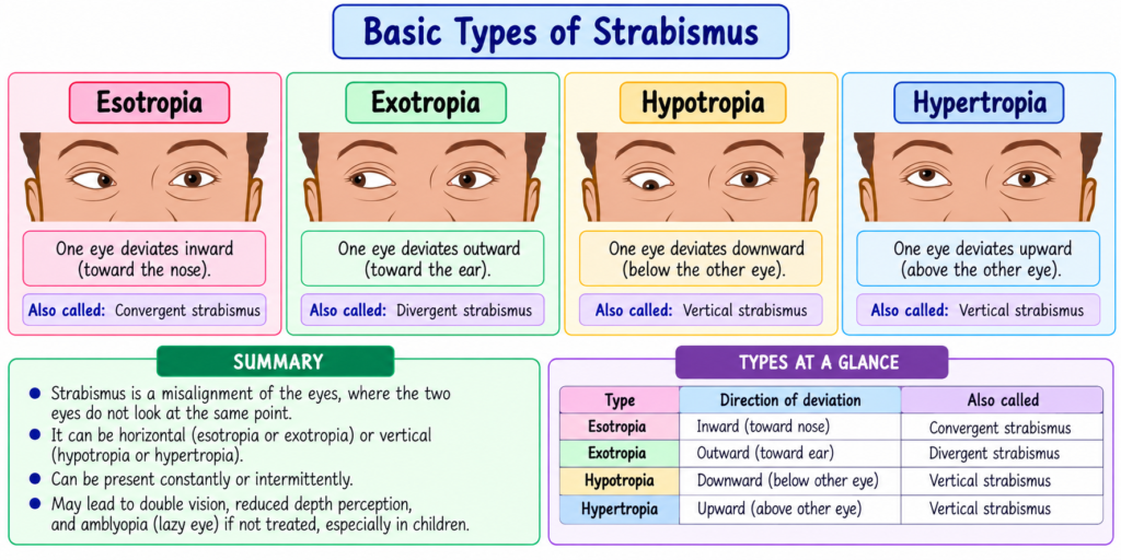
The basic types are shown in Fig. 52.10.
Esotropia
Esotropia: One or both eyes turn inward toward the nose.
Exotropia
Exotropia: One or both eyes turn outward.
Hypertropia
Hypertropia: One or both eyes turn upward.
Hypotropia
Hypotropia: One or both eyes turn downward.
Comparison of Strabismus Types
| Type | Direction |
|---|---|
| Esotropia | Inward |
| Exotropia | Outward |
| Hypertropia | Upward |
| Hypotropia | Downward |
Causes of Strabismus
Strabismus may result from abnormalities involving:
- Extraocular muscles.
- Nerves supplying these muscles.
- Brain centers controlling eye movement.
These abnormalities may be:
- Congenital.
- Acquired.
Acquired causes mentioned in the text include:
- Diabetes mellitus.
- Stroke.
- Brain tumors.
- Injuries.
Strabismus in Early Childhood
Strabismus in childhood may develop because of an abnormal setting of the fusion mechanism.
During early fixation:
- One eye may fix normally while the other fails.
- Or both may fix properly but not simultaneously.
Over time:
Abnormal fixation pattern
→ conjugate eye-movement pathways become abnormally set
→ normal fusion does not develop
Suppression of the Visual Image From a Repressed Eye
In some patients:
- The eyes alternate fixation.
In others:
- One eye is used continuously.
- The other becomes repressed.
- The repressed eye is not used for precise vision.
Its visual acuity may remain:
20/400 or less
Effect of Age
If the dominant eye later becomes blind:
- In adults:
- The repressed eye improves only slightly.
- In young children:
- Vision may improve much more.
This shows that visual acuity depends strongly on proper development of central nervous system synaptic connections from the eyes.
In the visual cortex:inal Educational Figure / Diagram
Eye-Movement Control
Visual target
→ visual cortex
→ pretectal area + superior colliculus
→ oculomotor nuclei
→ extraocular muscles
→ eye movement
At the same time:
Vestibular nuclei
→ medial longitudinal fasciculus
→ oculomotor system
Fixation Control
Frontal voluntary eye fields
→ unlock current fixation
→ move eyes toward new target
Occipital involuntary eye fields
→ lock eyes on target
Image drifts toward edge of fovea
→ corrective flick
→ image returns toward foveal center
Key Concept
The major eye-movement functions can be remembered as:
Find a target
→ voluntary fixation
Hold the target
→ involuntary fixation
Jump to another target
→ saccade
Follow a moving target
→ pursuit movement
For binocular vision:
Two retinal images
→ cortical comparison
→ ocular adjustment
→ fusion
Image disparity between the two eyes
→ stereopsis
→ depth perception
Clinical Importance / Clinical Correlation
- Bilateral damage to frontal voluntary fixation areas can make it difficult to shift fixation.
- Damage to involuntary fixation areas can impair the ability to maintain fixation.
- Destruction of the superior colliculi greatly reduces involuntary fixation and orienting responses.
- Proper fusion requires coordinated alignment of the eyes.
- Strabismus results from abnormal ocular alignment.
- It may involve abnormalities of:
- Extraocular muscles.
- Their nerves.
- Brain control centers.
- Early suppression of one eye can interfere with normal development of visual acuity.
- Long-term repression reduces normal neuronal connections from that eye to the visual cortex.
High-Yield / Exam Points
- Medial and lateral recti → side-to-side movement.
- Superior and inferior recti → upward and downward movement.
- Oblique muscles → rotational movements.
- Cranial nerve nuclei involved:
- III
- IV
- VI
- Medial longitudinal fasciculus connects important oculomotor pathways.
- Voluntary fixation → frontal premotor cortex.
- Involuntary fixation → secondary occipital visual areas.
- Tremor frequency → 30–80 cycles/sec.
- Saccades → approximately 2–3 jumps/sec.
- About 90% of the time is spent at fixation points.
- About 10% or less is spent during saccadic movement.
- Pursuit movement keeps the eyes fixed on a moving object.
- Superior colliculi orient the eyes, head, and body toward external disturbances.
- Stereopsis is important up to about 200 ft (61 m).
- Monocular depth clues:
- Image size.
- Motion parallax.
- Image blocking.
- Esotropia → inward.
- Exotropia → outward.
- Hypertropia → upward.
- Hypotropia → downward.
- A repressed eye may remain at 20/400 or less visual acuity.
Common Student Mistakes
- Do not confuse voluntary and involuntary fixation.
- Voluntary → moves fixation.
- Involuntary → maintains fixation.
- Do not confuse saccades with pursuit movements.
- Saccades → rapid jumps.
- Pursuit → smooth following of a moving object.
- Do not think the eyes remain completely motionless during fixation.
- Tremor, drift, and flicking movements continue.
- Do not think stereopsis is the only depth mechanism.
- Monocular clues also contribute.
- Do not confuse the types of strabismus.
- Esotropia → inward.
- Exotropia → outward.
- Hypertropia → upward.
- Hypotropia → downward.
Quick Revision
Eye muscles
Medial/lateral recti
→ side to side
Superior/inferior recti
→ up and down
Obliques
→ rotation
Fixation
Frontal cortex
→ voluntary fixation
→ move to new target
Occipital secondary visual areas
→ involuntary fixation
→ hold target
Fixation correction
Drift toward foveal edge
→ corrective flick
→ image returns centrally
Saccades
One fixation point
→ rapid jump
→ next fixation point
Pursuit
Moving target
→ eye movement gradually matches target
→ smooth following
Superior colliculi
External disturbance
→ eyes + head + body orient toward it
Fusion
Two misaligned retinal images
→ cortical signal
→ convergence/divergence/rotation
→ images align
Stereopsis
Binocular image disparity
→ judgment of distance
Strabismus
Misalignment of eyes
→ failure of normal fusion
Final Take-Home Concept / Summary
Eye movement depends on coordinated control of extraocular muscles, brain stem nuclei, cortical visual areas, vestibular centers, and the superior colliculi.
Voluntary fixation
→ finds a new target
Involuntary fixation
→ keeps the target on the fovea
Saccades
→ rapidly shift between fixation points
Pursuit movements
→ follow moving objects
The superior colliculi help orient the:
- Eyes.
- Head.
- Body.
toward visual, auditory, and somatic disturbances.
Normal binocular vision requires fusion:
Two retinal images
→ cortical comparison
→ ocular adjustment
→ fused image
Differences between the two retinal images produce:
→ stereopsis
→ depth perception
Strabismus is a failure of normal ocular alignment and fusion. When one eye is chronically repressed during development, normal visual acuity and cortical neuronal connections from that eye may fail to develop properly.
References / Sources
- Guyton and Hall Textbook of Medical Physiology, 15th Edition, Chapter 52.
