


Learning Objectives
After completing this lecture, students will be able to:
- Explain how the visual cortex detects contrast, lines, and borders.
- Differentiate simple cells, complex cells, and higher-order visual responses.
- Explain how color contrast and color constancy are analyzed.
- Describe the effects of loss of the primary visual cortex.
- Explain visual fields, perimetry, scotomata, and retinitis pigmentosa.
Introduction
The visual cortex does not simply respond equally to every illuminated part of a visual scene.
Instead, it is especially responsive to:
- Changes between light and dark areas.
- Borders and lines.
- Orientation of lines.
- Specific shapes.
- Color contrasts.
As visual information travels through the visual cortex, increasingly complex features of the image are identified.
The visual system also allows examination of the field of vision, which can help identify abnormalities affecting different parts of the retina or visual pathway.
Main Topic / Core Concept
Analysis of Contrasts in Visual Images
When a person looks at a blank wall:
- Only a few neurons in the primary visual cortex are stimulated.
- This remains true whether the wall is:
- Brightly illuminated.
- Weakly illuminated.
This shows that the primary visual cortex is not mainly concerned with uniform illumination.
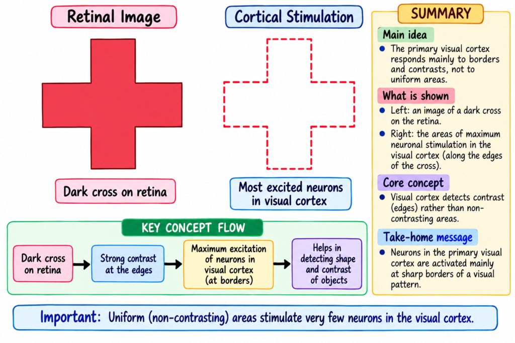
When a large solid cross is placed on the wall, the strongest cortical excitation occurs along its sharp borders, as demonstrated in Fig. 52.5.
Therefore:
- The primary visual cortex responds mainly to contrast in a visual image.
- Noncontrasting areas produce much less cortical stimulation.
Contrast: A difference between adjacent light and dark areas of the visual scene.
Why Borders Produce Strong Responses
Adjacent retinal receptors that are stimulated equally tend to inhibit one another.
Therefore:
Uniform illumination
→ neighboring receptors are similarly stimulated
→ mutual inhibition
→ weak cortical response
At a border:
Dark area meets light area
→ neighboring receptors are stimulated differently
→ mutual inhibition is reduced
→ strong neuronal response
The strength of neuronal stimulation depends on the gradient of contrast.
Contrast gradient: The degree of change between adjacent light and dark areas.
Therefore:
Sharper contrast
- greater difference between light and dark
→ greater neuronal stimulation
Detection of Line and Border Orientation — Simple Cells
The visual cortex detects:
- The presence of a line or border.
- The direction in which the line is oriented.
A line may be:
- Vertical.
- Horizontal.
- Inclined at another angle.
Specific cortical neurons respond to specific orientations.
These neurons are called simple cells.
Simple cells: Visual cortical neurons that respond to lines or borders with particular orientations.
- Simple cells are found mainly in layer IV of the primary visual cortex.
- A line in one direction activates one group of simple cells.
- Changing the direction of the line activates another group.
Concept
Vertical line
→ one group of simple cells
Horizontal line
→ different group of simple cells
Inclined line
→ another group of simple cells
Complex Cells
As visual information moves farther away from layer IV:
- Some neurons continue to respond to a line with a particular orientation.
- Their response is less dependent on the exact position of the line.
These neurons are called complex cells.
Complex cells: Visual cortical neurons that respond to a line of a particular orientation even when the line moves moderate distances laterally or vertically.
For example:
Same line orientation
- different location
→ same complex cells may still respond
Simple Cells vs Complex Cells
| Feature | Simple Cells | Complex Cells |
|---|---|---|
| Main feature detected | Line or border orientation | Line orientation |
| Position dependence | More position-specific | Less position-specific |
| Response when line moves | Different cells may respond | Same cells may continue responding |
| Main location described | Mainly layer IV | Farther from layer IV |
Detection of Lengths, Angles, and Other Shapes
Some neurons respond to even more specific features.
These neurons are present in:
- Outer layers of primary visual cortical columns.
- Some secondary visual areas.
They may respond only to:
- Lines of particular lengths.
- Particular angles.
- Specific shapes.
- Images with other special characteristics.
Therefore, as visual signals move farther through the analytical pathway:
Simple visual features
→ more complex visual characteristics
→ progressively greater analysis of the visual scene
Detection of Color
Color is analyzed in a way similar to line detection.
The visual cortex uses color contrast.
Color Constancy
Color constancy: The ability to continue interpreting an object’s color appropriately even when the color of the illuminating light changes.
The comparison of colors with white is believed to be especially important.
When the illuminating light changes:
Illuminating light changes
→ appearance of white also changes
→ brain makes an appropriate comparison
→ an object can still be interpreted as having its expected color
For example:
Changing illumination
→ light entering the eye from a red object changes
→ appropriate brain processing
→ object is still interpreted as red
Opponent Colors
Opponent colors: Contrasting colors that activate specific visual neurons in an opposing manner.
Color contrast analysis depends on opposing color relationships.
The text describes examples such as:
- Red versus green.
- Blue versus red.
- Green versus yellow.
It is presumed that:
- Initial color contrast details are detected by simple cells.
- More complicated color contrasts are detected by:
- Complex cells.
- Hypercomplex cells.
Mechanism / Pathway / Step-by-Step Explanation
Contrast Detection
Uniform visual field
→ neighboring retinal receptors stimulated similarly
→ mutual inhibition
→ little cortical excitation
Contrast border appears
→ light-dark stimulation becomes unequal
→ mutual inhibition is reduced at the border
→ cortical neurons are strongly stimulated
→ border becomes detectable
Greater light-dark difference
→ greater contrast gradient
→ stronger neuronal stimulation
Progressive Analysis of Visual Form
Visual border
→ simple cells detect orientation
→ complex cells detect similar orientation even when position changes
→ higher-order neurons detect lengths, angles, and other shapes
→ visual characteristics become progressively analyzed
Color Analysis
Different colors within visual scene
→ colors are compared with surrounding colors or white
→ opponent-color neurons respond
→ simple cells analyze initial color contrast
→ complex and hypercomplex cells analyze more complicated contrasts
Original Educational Diagram
Progressive cortical analysis of a visual image
Visual image
→ contrast borders detected
→ line orientation detected
→ position-independent line orientation detected
→ length / angle / shape analyzed
→ increasingly complex features of the visual scene are deciphered
Educational explanation:
The passage shows that visual analysis becomes progressively more complex as signals move through the cortical analytical pathway. The cortex first emphasizes contrast and borders and then identifies orientation, shape, and other visual characteristics.
Effect of Removing the Primary Visual Cortex
Removal of the primary visual cortex causes:
→ loss of conscious vision
→ blindness
However, some visual reactions may still occur subconsciously.
A person may sometimes respond to:
- Changes in light intensity.
- Movement in the visual scene.
- Rarely, some gross visual patterns.
Possible responses include:
- Turning the eyes.
- Turning the head.
- Avoidance.
These residual responses are believed to depend mainly on pathways traveling:
Optic tracts
→ superior colliculi
→ other parts of the older visual system
Key Concept
Primary visual cortex removed
→ conscious vision lost
Older visual pathways remain
→ some subconscious visual responses may persist
Fields of Vision and Perimetry
Field of Vision
Field of vision: The total visual area that can be seen by one eye at a particular moment.
It is divided into:
- Nasal field: Area seen toward the nasal side.
- Temporal field: Area seen toward the lateral side.
Perimetry
Perimetry: A method used to chart the field of vision of each eye and identify blindness in specific portions of the visual field.
During perimetry:
- One eye looks directly at a central point.
- The opposite eye is closed.
- A small light or object is moved through different parts of the visual field.
- The person reports:
- When the object is visible.
- When the object is not visible.
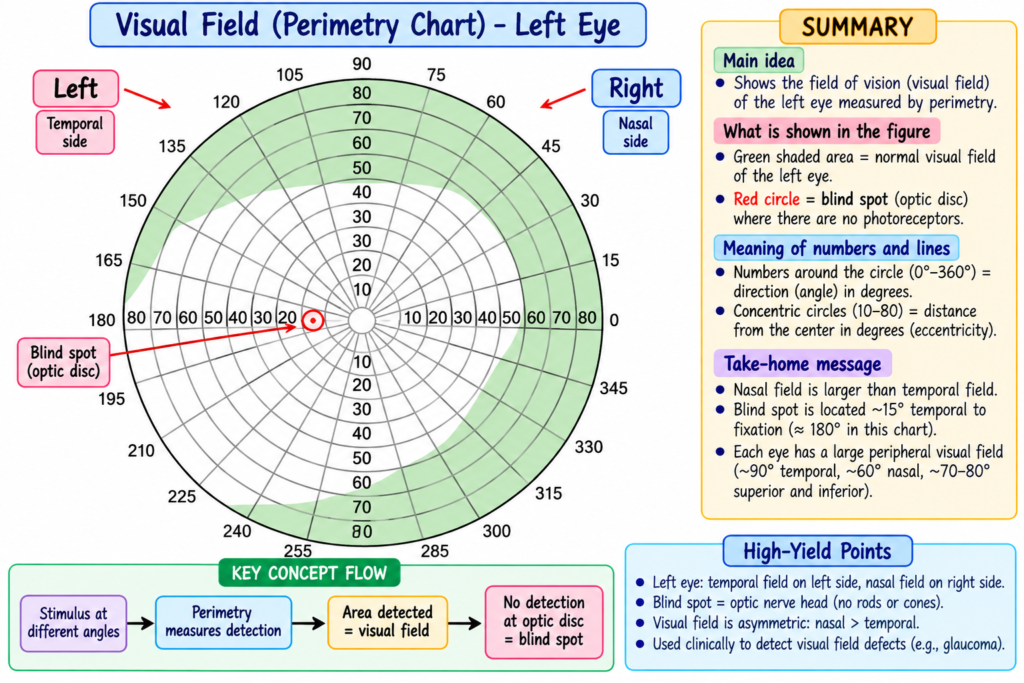
The visual field of the left eye is illustrated in Fig. 52.6.
Normal Blind Spot
A normal blind spot is present in every perimetry chart.
It occurs because:
- The optic disc contains no rods or cones.
Its location is approximately:
15° lateral to the central point of vision
Mechanism
Optic disc
→ no rods or cones
→ light falling there cannot be detected
→ normal blind spot
Abnormalities in the Fields of Vision
Scotomata
Scotoma: An abnormal blind spot located in a portion of the visual field other than the normal optic-disc blind spot.
Scotomata may result from:
- Optic nerve damage caused by glaucoma.
- Allergic reactions in the retina.
- Toxic conditions such as:
- Lead poisoning.
- Excessive use of tobacco.
Glaucoma: A condition described in the provided text as excessive fluid pressure within the eyeball that can damage the optic nerve.
Retinitis Pigmentosa
Retinitis pigmentosa: A condition in which parts of the retina degenerate and excessive melanin pigment is deposited in the degenerated areas.
The visual loss typically progresses as follows:
Peripheral field blindness first
→ visual loss gradually extends inward
→ central visual areas become progressively involved
Key Concept
The primary visual cortex is particularly designed to analyze differences within the visual scene, rather than uniform illumination.
Visual processing follows a progressive pattern:
Contrast
→ borders
→ orientation
→ position-independent orientation
→ lengths and angles
→ complex shapes and visual characteristics
Similarly:
Color contrast
→ opponent-color analysis
→ increasingly complex color processing
Clinical Importance / Clinical Correlation
- Destruction of the primary visual cortex causes loss of conscious vision.
- Some subconscious visual reactions may remain because older visual pathways continue to function.
- Perimetry can identify missing portions of the visual field.
- The normal blind spot is produced by the absence of rods and cones at the optic disc.
- Abnormal blind spots are called scotomata.
- Scotomata may occur with:
- Glaucoma-related optic nerve damage.
- Retinal allergic reactions.
- Lead poisoning.
- Excessive tobacco use.
- Perimetry can also help identify the visual-field changes of retinitis pigmentosa.
- In retinitis pigmentosa:
- Peripheral visual loss occurs first.
- Visual loss gradually approaches the central field.
High-Yield / Exam Points
- The primary visual cortex responds strongly to contrast borders, not uniform illumination.
- Sharper light-dark differences produce stronger cortical stimulation.
- Simple cells mainly detect orientation of lines and borders.
- Simple cells are found mainly in layer IV.
- Complex cells continue to respond to the same line orientation even when the line changes position.
- Higher-order neurons detect:
- Specific lengths.
- Angles.
- Shapes.
- Color analysis also depends on contrast.
- Comparison with white contributes to color constancy.
- Opponent colors activate specific cortical neurons.
- Removal of the primary visual cortex causes loss of conscious vision.
- Some subconscious visual reactions may survive through older visual pathways.
- Perimetry charts the visual field.
- Normal blind spot:
- Caused by absence of rods and cones at the optic disc.
- Approximately 15° lateral to central vision.
- An abnormal blind spot is called a scotoma.
- Retinitis pigmentosa usually affects peripheral vision first.
Common Student Mistakes
- Do not think uniform bright light produces maximal activity in the primary visual cortex.
- Sharp contrast borders produce much stronger responses.
- Do not confuse simple and complex cells.
- Simple cells → orientation and position are important.
- Complex cells → orientation remains important even when position changes.
- Do not think visual analysis stops after line detection.
- Higher cortical neurons analyze lengths, angles, shapes, and other features.
- Do not confuse the normal blind spot with a scotoma.
- Normal blind spot → optic disc.
- Scotoma → abnormal blind area elsewhere in the visual field.
- Do not think removal of the primary visual cortex eliminates every possible visual response.
- Conscious vision is lost, but some subconscious responses may remain through older visual pathways.
Quick Revision
Blank uniform wall
→ little primary visual cortical stimulation
Sharp border
→ strong cortical stimulation
Sharper contrast
→ stronger response
Simple cell
→ detects line orientation
Complex cell
→ detects orientation even when line position changes
Higher-order neurons
→ length + angles + shapes
Color contrast
→ opponent-color neurons
Primary visual cortex removed
→ conscious blindness
→ some subconscious responses may remain
Perimetry
→ maps visual field
Optic disc
→ no rods/cones
→ normal blind spot
Scotoma
→ abnormal blind spot
Retinitis pigmentosa
→ peripheral blindness first
→ gradually progresses centrally
Final Take-Home Concept / Summary
The primary visual cortex responds mainly to differences and borders within a visual scene rather than to uniform illumination.
Visual analysis becomes progressively more complex:
Contrast → line orientation → position-independent orientation → length and angle → complex shape
Simple cells mainly detect line orientation, whereas complex cells can respond to the same orientation even when the line changes position.
Color is also analyzed through contrast, including relationships between opponent colors and comparisons with white that contribute to color constancy.
Loss of the primary visual cortex causes loss of conscious vision, although some subconscious responses can remain through older visual pathways.
The visual field can be examined by perimetry. The normal blind spot results from the absence of photoreceptors at the optic disc, whereas abnormal blind areas are called scotomata. Retinitis pigmentosa typically causes peripheral visual-field loss before progressively involving more central areas.
Final concept:
Visual cortex
→ detects contrast first
→ analyzes orientation and shape
→ analyzes increasingly complex visual information
Perimetry
→ maps the visual field
→ identifies normal and abnormal blind areas
References / Sources
- Guyton and Hall Textbook of Medical Physiology, 15th Edition, Chapter 52.
