


Learning Objectives
After studying this lecture, students will be able to:
- Describe the organization of the primary and secondary visual cortex.
- Explain the six layers and vertical columns of the primary visual cortex.
- Differentiate the M and P visual pathways.
- Explain how the cortex processes color, binocular vision, position, motion, and visual detail.
Introduction
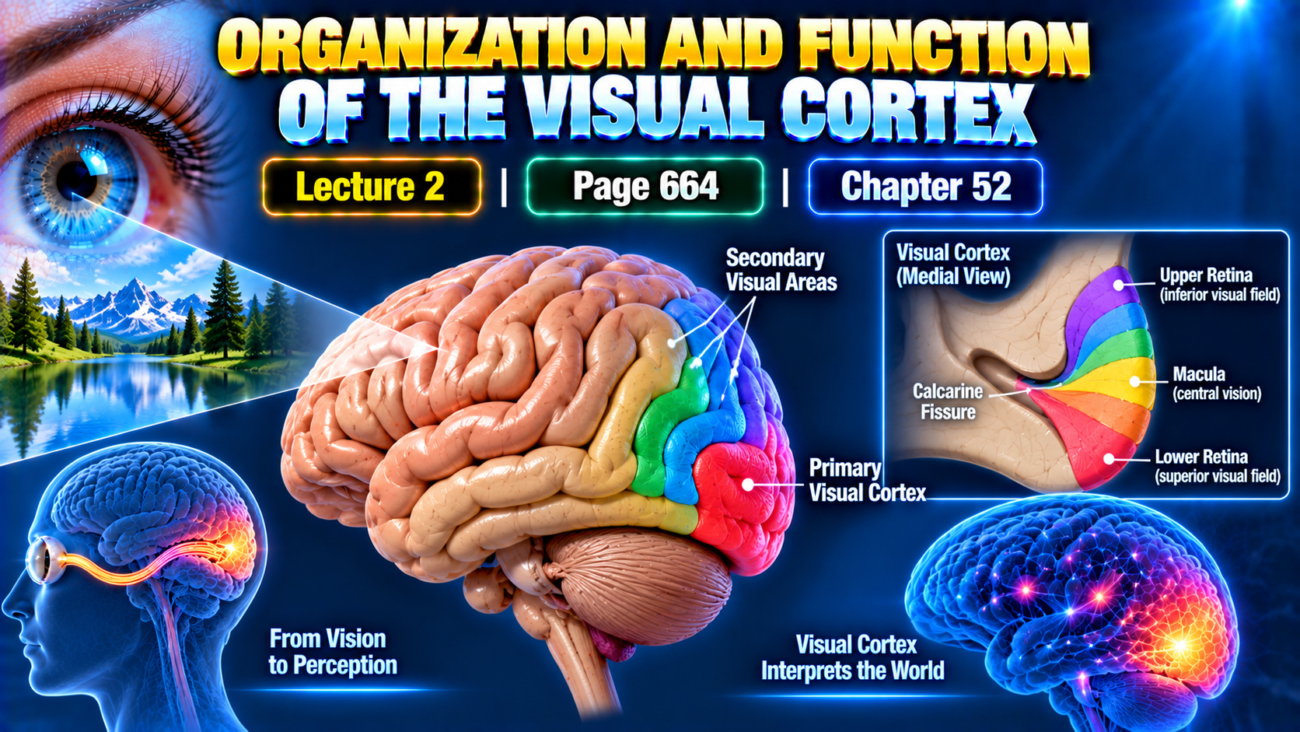
- The visual cortex is located mainly on the medial aspect of the occipital lobes.
- It receives and analyzes visual information coming from the eyes.
- Like other sensory cortical systems, it is divided into:
- Primary visual cortex
- Secondary visual areas
- These regions work together to analyze different components of the visual image.
Figs. 52.2 and 52.3 show the organization of the visual cortex.
Primary Visual Cortex
Definition
- The primary visual cortex is the cortical area where direct visual signals from the eyes terminate.
- It is also called:
- Visual area I
- V1
- Striate cortex
Location
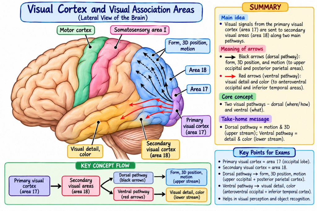
- It lies in the calcarine fissure area.
- It extends forward from the occipital pole.
- It is located on the medial aspect of each occipital cortex.
Fig. 52.2 shows the primary visual cortex and its retinal representation.
Representation of the Retina
- Signals from the macular area of the retina terminate near the occipital pole.
- Signals from the peripheral retina terminate farther anteriorly.
- These peripheral retinal signals are arranged in concentric half-circles along the calcarine fissure.
- The upper retina is represented superiorly.
- The lower retina is represented inferiorly.
Large Cortical Representation of the Macula
- A very large area of the primary visual cortex represents the macula.
- The fovea sends its visual signals to this region.
- The fovea provides the highest degree of visual acuity.
- Compared with peripheral retinal areas, the fovea has several hundred times greater cortical representation when considered in relation to retinal area.
Why Is It Called the Striate Cortex?
- The primary visual cortex has a grossly striated appearance.
- Therefore, it is also called the striate cortex.
Secondary Visual Areas
Definition
- The secondary visual areas are also called visual association areas.
- They receive visual information after its initial processing in the primary visual cortex.
- Their main role is further analysis of the meaning of visual information.
Location
They lie:
- Lateral to the primary visual cortex
- Anterior to it
- Superior to it
- Inferior to it
Many of these areas extend onto the lateral surfaces of the:
- Occipital cortex
- Parietal cortex
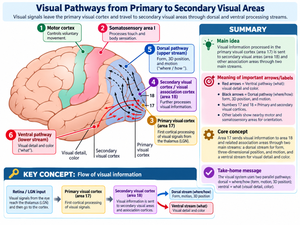
Fig. 52.3 shows the distribution of these secondary visual areas.
Visual Area II
- Brodmann area 18 surrounds the primary visual cortex.
- Almost all signals leaving the primary visual cortex pass next to this area.
- Therefore, Brodmann area 18 is called:
- Visual area II
- V2
Other Visual Areas
Other secondary visual areas include:
- V3
- V4
- Additional higher visual areas
There are more than a dozen specialized visual areas.
Core Concept
Visual information is not analyzed all at once.
Instead:
Primary visual cortex → Secondary visual areas → Progressive analysis of different features of the visual image
Different parts of the image are progressively separated, examined, and interpreted.
The Primary Visual Cortex Has Six Major Layers
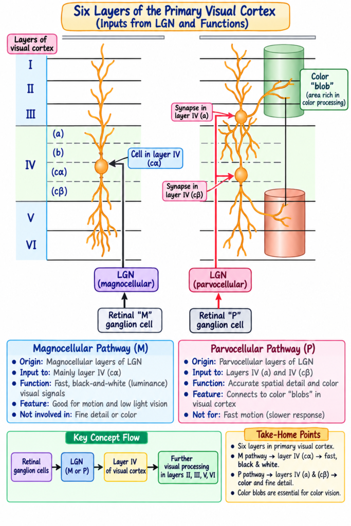
- Like most of the cerebral cortex, the primary visual cortex contains six distinct layers.
Fig. 52.4 shows these cortical layers and their organization.
Layer IV
- Visual fibers from the geniculocalcarine pathway terminate mainly in layer IV.
- Layer IV itself is divided into subdivisions.
Two important visual signal systems terminate in different parts of layer IV:
- M pathway
- P pathway
M Ganglion Cell Pathway
M Ganglion Cells
- Signals from M retinal ganglion cells are conducted rapidly.
- These signals terminate mainly in layer IVcα.
From layer IVcα:
M signals → Spread vertically upward toward the cortical surface + downward toward deeper cortical layers
Main Functional Features
The M pathway mainly carries information related to:
- Rapid visual signals
- Position of objects
- Gross form
- Motion
- Black-and-white visual information
P Ganglion Cell Pathway
P Ganglion Cells
- Signals from medium-sized optic nerve fibers originate from P retinal ganglion cells.
- These signals also terminate in layer IV.
- However, they terminate at different locations from the M pathway.
They mainly terminate in:
- Layer IVa
- Layer IVcβ
From there:
P signals → Travel vertically toward superficial layers + deeper cortical layers
Main Functional Features
The P pathway carries:
- Accurate point-to-point vision
- Fine visual detail
- Color vision
Vertical Neuronal Columns in the Visual Cortex
Definition
- The visual cortex contains several million vertical neuronal columns.
- Each column represents a functional unit.
Size
- Each column is approximately 30–50 micrometers in diameter.
- Each column may contain about 1000 or more neurons.
Functional Organization
After optic signals arrive in layer IV:
Layer IV → Signals spread upward and downward through the vertical column → Further visual processing occurs
This organization helps analyze separate components of visual information at successive processing stations.
Signals Moving Toward Superficial Layers
Signals passing outward reach:
- Layer I
- Layer II
- Layer III
These signals eventually travel short distances laterally within the cortex.
Signals Moving Toward Deeper Layers
Signals passing inward reach:
- Layer V
- Layer VI
Neurons in these layers transmit signals over much greater distances.
Color Blobs in the Visual Cortex
Definition
- Color blobs are specialized column-like regions found among visual cortical columns.
They are present in:
- Primary visual cortex
- Some secondary visual areas
Function
- They receive lateral signals from neighboring visual columns.
- They are specifically activated by color signals.
- They are therefore considered important areas for deciphering color information.
Key Flow
Color signals → Visual columns → Color blobs → Color analysis
Interaction of Visual Signals From the Two Eyes
- Visual information from the two eyes remains separated in the lateral geniculate nucleus.
- These signals are still separate when they reach layer IV of the primary visual cortex.
Alternating Cortical Stripes
- Layer IV contains alternating stripes of neuronal columns.
- Each stripe is approximately 0.5 mm wide.
- Signals from one eye enter every other stripe.
- Signals from the opposite eye enter the alternating stripes.
Image Registration
The visual cortex compares corresponding retinal images from both eyes.
It determines whether:
Image from one eye + Image from the other eye → Correspond correctly
This matching is described as the images being “in register.”
Fusion of the Two Images
Information about image registration helps adjust the direction of the two eyes.
This allows:
Alignment of both eyes → Corresponding images match → Visual images fuse
Stereopsis
- The degree of matching between the two retinal images also provides information about the distance of objects.
- This mechanism is called stereopsis.
Two Major Pathways for Analysis of Visual Information
After visual information leaves the primary visual cortex, it enters two major analytical pathways.
These are:
- Fast position and motion pathway
- Accurate visual detail and color pathway
Fast Position and Motion Pathway
This pathway analyzes:
- Three-dimensional position of objects
- Gross physical form
- Motion
It determines:
- Where an object is
- Whether the object is moving
Pathway
Primary visual cortex → Posterior midtemporal region → Broad occipitoparietal cortex
At the anterior border of the parietal cortex:
- Visual signals overlap with signals from posterior somatic association areas.
- These somatic areas also analyze three-dimensional aspects of sensory information.
Main Input
This pathway mainly receives information from:
M retinal ganglion cells → Large M optic nerve fibers → Rapid visual signals
Information Carried
- Position
- Gross form
- Motion
- Rapid information
- Black-and-white information
- No color information
Visual Detail and Color Pathway
This pathway passes from the primary visual cortex into secondary visual areas of the:
- Inferior occipital cortex
- Ventral occipital cortex
- Medial occipital cortex
- Temporal cortex
Main Functions
It analyzes:
- Fine visual detail
- Color
- Letters
- Reading
- Surface texture
- Detailed colors of objects
- Identity and meaning of objects
Conceptual Flow
Primary visual cortex → Inferior/ventral visual areas → Fine detail + Color analysis → Recognition of the object and its meaning
Key Concept
Visual information is progressively analyzed rather than interpreted by one cortical area alone.
Eyes → Primary visual cortex → Secondary visual areas → Specialized analysis
The two major processing streams can be remembered as:
M pathway → Where is it? Is it moving?
P-related detailed pathway → What is it? What color and detail does it have?
Clinical Importance / Functional Importance
The organization of the visual cortex explains several important visual functions described in the provided text:
- The large cortical representation of the fovea supports very high visual acuity.
- Interaction between signals from both eyes allows proper image fusion.
- Comparison of images from the two eyes contributes to stereopsis and distance perception.
- The position-motion pathway helps determine where an object is and whether it is moving.
- The visual-detail pathway allows recognition of:
- Letters
- Surface texture
- Color
- Objects
- Meaning of visual information
High-Yield Points
- Primary visual cortex = V1 = visual area I = striate cortex.
- It is located mainly around the calcarine fissure.
- Macular and foveal vision has a very large cortical representation.
- Brodmann area 18 = V2.
- Geniculocalcarine fibers mainly terminate in layer IV.
- M ganglion signals terminate mainly in IVcα.
- P ganglion signals terminate mainly in IVa and IVcβ.
- M pathway = rapid signals, position, gross form, motion, black and white.
- P pathway = accurate point-to-point vision and color.
- Color blobs participate in color analysis.
- Alternating cortical stripes receive signals from the two separate eyes.
- Matching information from both eyes contributes to stereopsis.
Common Student Mistakes
- Do not confuse V1 with V2.
- V1 = primary visual cortex.
- V2 = Brodmann area 18.
- Do not confuse the M pathway with the P pathway.
- M = rapid, position and motion.
- P = fine detail and color.
- Do not assume the whole retina has equal cortical representation.
- The fovea has a much larger representation.
- Do not think signals from both eyes immediately mix completely.
- They remain separated when they first arrive in layer IV.
- Do not confuse the two major visual analysis pathways.
- One determines mainly where an object is and whether it moves.
- The other helps determine what the object is.
Quick Revision
Visual Cortex
→ Primary visual cortex
→ Secondary visual areas
Primary Visual Cortex
→ Calcarine fissure
→ V1
→ Striate cortex
→ Direct visual signals arrive here
Macula/Fovea
→ Large cortical representation
→ Highest visual acuity
Layer IV
→ Main receiving layer
M cells
→ IVcα
→ Rapid signals
→ Position + Motion + Gross form
→ Black and white
P cells
→ IVa + IVcβ
→ Accurate vision
→ Fine detail + Color
Vertical columns
→ Functional processing units
Color blobs
→ Color analysis
Signals from both eyes
→ Alternating cortical columns
→ Image matching
→ Fusion
→ Stereopsis
Two major visual pathways
→ Position/Motion pathway = Where?
→ Detail/Color pathway = What?
Final Take-Home Concept
The visual cortex works as an organized processing system.
Visual signals first reach the primary visual cortex, where the retinal image is precisely represented.
The information then passes through specialized cortical layers, vertical columns, color-processing regions, and secondary visual areas.
Two major processing systems then analyze different features:
M-related pathway → Position + Gross form + Motion
Detailed visual pathway → Fine detail + Color + Object recognition
Together, these cortical systems allow the brain to determine:
Where an object is → Whether it is moving → What it looks like → What it means
Reference Guyton and Hall Textbook of Medical Physiology 15th Edition, page. 664 Chapter 52
