


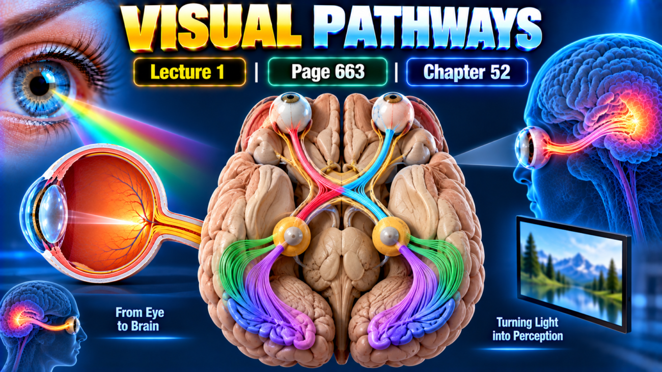
Learning Objectives
After studying this topic, students will be able to:
- Trace the main visual pathway from the retina to the visual cortex.
- Explain the older and newer visual pathways.
- Describe the functions of the dorsal lateral geniculate nucleus.
- Compare magnocellular and parvocellular pathways.
Introduction
- Visual information from both retinas travels through a highly organized pathway toward the visual cortex.
- Some optic nerve fibers cross at the optic chiasm, while others remain on the same side.
- The main conscious visual pathway passes through the dorsal lateral geniculate nucleus of the thalamus before reaching the primary visual cortex.
- Other visual fibers travel to older brain areas concerned with:
- Circadian rhythms
- Pupillary light reflexes
- Eye movements
- Behavioral functions
- The dorsal lateral geniculate nucleus does more than relay signals.
- It also:
- Preserves accurate spatial information.
- Keeps signals from the two eyes separate.
- Controls how much visual information reaches the visual cortex.
PRINCIPAL VISUAL PATHWAY
Visual pathway: The neural route by which visual signals travel from the retina toward the brain and visual cortex.
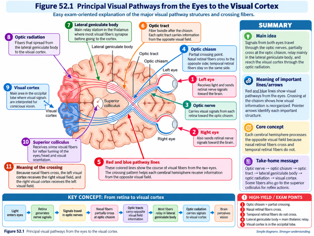
The principal visual pathways are shown in Fig. 52.1.
Main Sequence
Retina
→ optic nerve
→ optic chiasm
→ optic tract
→ dorsal lateral geniculate nucleus
→ optic radiation
→ primary visual cortex
Optic Nerves
Optic nerve: The pathway through which visual nerve signals leave the retina.
- Visual nerve signals leave both retinas through the optic nerves.
- The optic nerves travel toward the optic chiasm.
Optic Chiasm
Optic chiasm: The point where optic nerve fibers from the nasal halves of the retinas cross to the opposite side.
At the optic chiasm:
- Fibers from the nasal half of each retina cross to the opposite side.
- Fibers from the temporal retina remain on their side.
- After the crossing:
- Nasal retinal fibers join fibers from the opposite temporal retina.
- Together, they form the optic tract.
Key Concept
Nasal retinal fibers
→ cross at optic chiasm
Temporal retinal fibers
→ remain uncrossed
Then:
Crossed nasal fibers + opposite temporal fibers
→ optic Tracts
Optic tract: The visual pathway formed after fibers pass through the optic chiasm.
- Each optic tract contains fibers from both eyes.
- Its fibers travel toward the thalamus.
- They synapse mainly in the dorsal lateral geniculate nucleus.
Dorsal Lateral Geniculate Nucleus
Dorsal lateral geniculate nucleus: A thalamic nucleus that receives optic tract signals and relays them toward the visual cortex.
- Optic tract fibers synapse in this nucleus.
- From there, visual signals travel through the geniculocalcarine fibers.
Optic Radiation
Optic radiation: Fibers carrying visual signals from the dorsal lateral geniculate nucleus to the primary visual cortex.
It is also called the:
- Geniculocalcarine tract
Signal Flow
Dorsal lateral geniculate nucleus
→ optic radiation
→ primary visual cortex
Primary Visual Cortex
Primary visual cortex: The cortical area receiving the main visual signals from the lateral geniculate nucleus.
- It lies in the:
- Calcarine fissure area
- Medial occipital lobe
Complete Main Pathway
Retina
→ optic nerve
→ optic chiasm
→ optic tract
→ dorsal lateral geniculate nucleus
→ optic radiation
→ primary visual cortex in the medial occipital lobe
VISUAL FIBERS TO OLDER BRAIN AREAS
Not all visual fibers travel directly toward the visual cortex.
Some pass to older areas of the brain.
Suprachiasmatic Nucleus
Suprachiasmatic nucleus: A hypothalamic nucleus involved in controlling circadian rhythms.
Pathway
Optic tract
→ suprachiasmatic nucleus of hypothalamus
Function
- Presumably helps control circadian rhythms.
- These rhythms synchronize physiological changes of the body with:
- Night
- Day
Circadian rhythm: A physiological rhythm synchronized with the day-night cycle.
Pretectal Nuclei
Pretectal nuclei: Midbrain nuclei involved in reflex eye responses and the pupillary light reflex.
Pathway
Visual fibers
→ pretectal nuclei in midbrain
Functions
- Produce reflex movements of the eyes.
- Help the eyes focus on important objects.
- Activate the pupillary light reflex.
Pupillary light reflex: The reflex response of the pupil to light.
Superior Colliculus
Superior colliculus: A brain region involved in rapid directional movements of the eyes.
Pathway
Visual fibers
→ superior colliculus
Function
- Controls rapid directional movements of both eyes.
Ventral Lateral Geniculate Nucleus and Basal Brain Regions
Some visual fibers pass to:
- Ventral lateral geniculate nucleus of the thalamus.
- Surrounding basal regions of the brain.
Function
- Presumably help control certain behavioral functions of the body.
OLD AND NEW VISUAL SYSTEMS
The visual pathways can be divided roughly into:
- An old visual system
- A new visual system
| Feature | Old Visual System | New Visual System |
|---|---|---|
| Main destination | Midbrain and base of forebrain | Visual cortex |
| Major role | Reflex and older visual functions | Conscious vision |
| Important areas | Suprachiasmatic nucleus, pretectal nuclei, superior colliculus, basal regions | Occipital visual cortex |
| In humans | Limited compared with cortical system | Responsible for nearly all conscious visual perception |
| In primitive animals | Can detect visual form | Less dominant compared with mammals |
Old Visual System
Old visual system: Visual pathways projecting mainly to the midbrain and base of the forebrain.
- These pathways participate in:
- Circadian control
- Reflex eye movements
- Pupillary responses
- Rapid directional eye movements
- Some behavioral functions
- In many primitive animals:
- Even visual form is detected by this older system.
- The superior colliculus performs a role similar to that of the visual cortex in mammals.
New Visual System
New visual system: The pathway that directly transmits visual signals toward the visual cortex.
- In humans, this system is responsible for the perception of virtually all:
- Visual form
- Colors
- Other aspects of conscious vision
Key Concept
Old visual system
→ mainly reflex and older visual functions
New visual system
→ conscious visual perception
FUNCTION OF THE DORSAL LATERAL GENICULATE NUCLEUS OF THE THALAMUS
Dorsal lateral geniculate nucleus: The main thalamic relay nucleus of the newer visual pathway.
- It is located at the dorsal end of the thalamus.
- It is also called the lateral geniculate body.
- Optic nerve fibers of the new visual system terminate here.
The nucleus has two major functions:
- Accurate relay of visual signals.
- Gating of visual transmission toward the cortex.
RELAY OF VISUAL INFORMATION
The first major function is to relay visual information.
Pathway
Optic tract
→ dorsal lateral geniculate nucleus
→ optic radiation
→ visual cortex
- This relay is extremely accurate.
- It provides exact point-to-point transmission.
- Spatial relationships are preserved with a high degree of fidelity from:
- Retina
- Through the thalamus
- To the visual cortex
Spatial fidelity: Accurate preservation of the location and arrangement of visual information during transmission.
Key Concept
A specific retinal point
→ corresponding geniculate neuron
→ corresponding visual cortical point
Therefore:
Retinal spatial organization is preserved.
SEPARATION OF SIGNALS FROM THE TWO EYES
After the optic chiasm:
- About half the fibers in each optic tract come from one eye.
- About half come from the other eye.
- These fibers represent corresponding retinal points.
However:
- Signals from the two eyes remain separated in the dorsal lateral geniculate nucleus.
Six Layers of the Dorsal Lateral Geniculate Nucleus
The nucleus contains six layers.
Layers II, III, and V
- Receive signals from the lateral half of the ipsilateral retina.
Ipsilateral: On the same side.
Layers I, IV, and VI
- Receive signals from the medial half of the retina of the opposite eye.
Key Concept
Some layers
→ receive signals from the same-side retina
Other layers
→ receive signals from the opposite-side retina
Yet:
- Corresponding retinal areas are represented in paired layers.
- Neurons representing corresponding points are positioned over one another.
- Parallel organization continues toward the visual cortex.
GATING OF VISUAL SIGNALS
The second major function of the dorsal lateral geniculate nucleus is gating.
Gating: Controlling how much visual information is allowed to pass to the visual cortex.
- Not every incoming visual signal is passed equally.
- The lateral geniculate nucleus can control transmission through selected regions.
Sources of Gating Control
Two major sources provide gating signals.
Corticofugal Fibers
Corticofugal fibers: Fibers traveling backward from the visual cortex toward the lateral geniculate nucleus.
Pathway
Primary visual cortex
→ backward fibers
→ lateral geniculate nucleus
Reticular Areas of the Mesencephalon
A second source comes from:
- Reticular areas of the mesencephalon.
Mesencephalon: The midbrain.
Effect of Gating Signals
- Both sources are inhibitory.
- When activated, they can reduce or stop transmission through selected parts of the dorsal lateral geniculate nucleus.
- These mechanisms help highlight the visual information that is allowed to pass onward.
Key Concept
Incoming visual signals
→ lateral geniculate nucleus
Inhibitory gating control
→ selected signals reduced
Important allowed information
→ continues toward visual cortex
MAGNOCELLULAR AND PARVOCELLULAR LAYERS
The dorsal lateral geniculate nucleus can also be divided according to cell size and input.
It contains:
- Magnocellular layers
- Parvocellular layers
MAGNOCELLULAR LAYERS
Magnocellular layers: Layers I and II of the dorsal lateral geniculate nucleus containing large neurons.
- Layers I and II are magnocellular.
- Their neurons are large.
- They receive input almost entirely from M retinal ganglion cells.
Functional Features
- Rapidly conducting pathway.
- Carries information quickly toward the visual cortex.
- Color blind.
- Transmits mainly black-and-white information.
- Point-to-point spatial transmission is relatively poor.
Why Spatial Accuracy Is Poorer
- There are relatively few M ganglion cells.
- Their dendrites spread widely across the retina.
Therefore:
Wide retinal sampling
→ less precise point-to-point localization
PARVOCELLULAR LAYERS
Parvocellular layers: Layers III through VI of the dorsal lateral geniculate nucleus containing many small to medium-sized neurons.
- Layers III to VI are parvocellular.
- They contain large numbers of:
- Small neurons
- Medium-sized neurons
- Their input comes almost entirely from P retinal ganglion cells.
Functional Features
- Transmit color information.
- Provide accurate point-to-point spatial information.
- Conduct signals at a moderate velocity.
- Their conduction is slower than the magnocellular pathway.
COMPARISON OF MAGNOCELLULAR AND PARVOCELLULAR PATHWAYS
| Feature | Magnocellular System | Parvocellular System |
|---|---|---|
| LGN layers | I and II | III through VI |
| Cell size | Large neurons | Small to medium-sized neurons |
| Main retinal input | M ganglion cells | P ganglion cells |
| Conduction speed | Rapid | Moderate |
| Color information | No | Yes |
| Main visual information | Black and white | Color |
| Point-to-point accuracy | Poorer | Accurate |
| Retinal ganglion distribution | Fewer M cells with widely spreading dendrites | P cells provide more precise spatial information |
KEY CONCEPT
The main visual pathway can be remembered as:
Retina
→ optic nerve
→ optic chiasm
→ optic tract
→ lateral geniculate nucleus
→ optic radiation
→ visual cortex
At the optic chiasm:
Nasal retinal fibers
→ cross
Temporal retinal fibers
→ remain on the same side
The lateral geniculate nucleus then:
- Relays signals accurately.
- Keeps signals from the two eyes separated.
- Controls how much visual information reaches the cortex.
- Separates information into magnocellular and parvocellular pathways.
CLINICAL / FUNCTIONAL IMPORTANCE
- Crossing of nasal retinal fibers allows visual information from the two eyes to be organized appropriately after the optic chiasm.
- The dorsal lateral geniculate nucleus preserves precise spatial relationships between the retina and visual cortex.
- Its gating function controls which visual signals are emphasized.
- Older visual pathways help control:
- Pupillary light reflexes
- Reflex eye movements
- Directional eye movements
- Circadian rhythms
- The newer cortical pathway is responsible for almost all conscious visual perception in humans.
- Magnocellular pathways provide rapid black-and-white information.
- Parvocellular pathways provide detailed spatial and color information.
HIGH-YIELD POINTS
- Nasal retinal fibers cross at the optic chiasm.
- Temporal retinal fibers remain on the same side.
- The major conscious pathway is:
Retina
→ optic nerve
→ optic chiasm
→ optic tract
→ dorsal lateral geniculate nucleus
→ optic radiation
→ primary visual cortex
- Primary visual cortex lies in the calcarine fissure region of the medial occipital lobe.
- Suprachiasmatic nucleus:
- Circadian rhythms.
- Pretectal nuclei:
- Reflex eye focusing.
- Pupillary light reflex.
- Superior colliculus:
- Rapid directional eye movements.
- Older visual system:
- Midbrain and forebrain-base pathways.
- Newer visual system:
- Visual cortex.
- Conscious form and color vision.
- Dorsal lateral geniculate nucleus:
- Accurate relay.
- Point-to-point spatial transmission.
- Keeps signals from the two eyes separate.
- Gates transmission to the cortex.
- The nucleus contains six layers.
- Layers I and II:
- Magnocellular.
- Layers III through VI:
- Parvocellular.
- Magnocellular pathway:
- Rapid
- Black and white
- Poorer spatial accuracy
- Parvocellular pathway:
- Color
- Accurate spatial detail
- Moderate conduction speed
COMMON STUDENT MISTAKES
- Do not think all optic nerve fibers cross at the optic chiasm.
- Only fibers from the nasal retinal halves cross.
- Do not confuse the optic tract with the optic nerve.
- Optic nerve is before the chiasm.
- Optic tract is after the chiasm.
- Do not think both eyes’ signals mix completely inside the lateral geniculate nucleus.
- They remain separated in different layers.
- Do not confuse old and new visual systems.
- Old system → midbrain and basal forebrain.
- New system → visual cortex.
- Do not confuse magnocellular with parvocellular pathways.
- Magnocellular → fast, black and white.
- Parvocellular → color and precise detail.
- Do not assume the lateral geniculate nucleus is only a passive relay.
- It also gates visual transmission.
QUICK REVISION
Main Visual Pathway
Retina
→ optic nerve
→ optic chiasm
→ optic tract
→ lateral geniculate nucleus
→ optic radiation
→ visual cortex
Chiasm
Nasal retina
→ crosses
Temporal retina
→ remains uncrossed
Older Visual Pathways
Suprachiasmatic nucleus
→ circadian rhythms
Pretectal nuclei
→ pupillary light reflex + reflex eye movements
Superior colliculus
→ rapid directional eye movements
Basal brain regions
→ behavioral functions
New Visual Pathway
Lateral geniculate nucleus
→ optic radiation
→ visual cortex
Function:
→ conscious visual form
→ color
→ other conscious vision
Lateral Geniculate Functions
Visual signal relay
→ accurate point-to-point transmission
Eye signals
→ remain separated
Gating
→ controls how much information reaches visual cortex
Magnocellular
Layers I and II
→ M ganglion cells
→ fast
→ black and white
→ poorer spatial precision
Parvocellular
Layers III–VI
→ P ganglion cells
→ color
→ accurate point-to-point information
→ moderate speed
CONCEPTUAL SUMMARY
Visual information begins in the retina and travels toward the brain through an organized pathway.
Retina
→ optic nerve
→ optic chiasm
At the chiasm:
Nasal retinal fibers
→ cross to the opposite side
Temporal retinal fibers
→ remain on the same side
These fibers combine to form:
→ optic tracts
Optic tract fibers then travel mainly to:
→ dorsal lateral geniculate nucleus of the thalamus
From there:
→ optic radiation
→ primary visual cortex in the medial occipital lobe
Some visual fibers also travel to older brain regions.
Suprachiasmatic nucleus
→ circadian rhythm control
Pretectal nuclei
→ pupillary and reflex eye responses
Superior colliculus
→ rapid directional eye movements
Basal brain regions
→ some behavioral functions
Therefore, the visual system can be divided into:
Old system
→ mainly reflex and older visual functions
New system
→ conscious vision through the visual cortex
The dorsal lateral geniculate nucleus has two major roles.
First:
Retinal signal
→ precise point-to-point relay
→ visual cortex
Second:
Incoming signal
→ gating mechanisms
→ selected information allowed to pass
The nucleus also contains separate magnocellular and parvocellular pathways.
Magnocellular system
M ganglion input
→ fast conduction
→ black-and-white information
→ poorer spatial precision
Parvocellular system
P ganglion input
→ moderate conduction
→ color information
→ precise spatial transmission
Final Take-Home Concept
Nasal retinal fibers cross; temporal retinal fibers do not.
The lateral geniculate nucleus is both a relay and a gate.
Magnocellular pathway = fast, black-and-white information.
Parvocellular pathway = color and precise spatial detail.
The visual cortex is the major center for conscious vision in humans.
Reference Guyton and Hall Textbook of Medical Physiology 15th Edition, page. 663 Chapter52
