Figure: Fig. 39.4 and Fig. 39.5
- Hydrostatic pressure is the pressure produced by the weight of blood inside the blood vessels.
- In a standing person, hydrostatic pressure affects blood flow in the lungs.
- The same effect occurs in the lungs as in the rest of the body, but to a lesser extent.
Hydrostatic Pressure Difference in the Lungs
- In an upright adult, the lowest part of the lungs is about 30 cm below the highest part.
- This height difference produces a hydrostatic pressure difference of about 23 mm Hg.
- Of this pressure difference:
- About 15 mm Hg is above the level of the heart.
- About 8 mm Hg is below the level of the heart.
Easy Concept
Think of the lungs as a 30 cm tall water tank.
Top of Lung
      │
      │ 15 mm Hg Lower
      │
Heart Level
      │
      │ 8 mm Hg Higher
      │
Bottom of Lung
Concept:
- Blood above the heart has lower pressure.
- Blood below the heart has higher pressure.
- This happens because blood has weight (hydrostatic pressure).
Pulmonary Arterial Pressure at Different Levels
- At the top of the lung, the pulmonary arterial pressure is about 15 mm Hg lower than at the level of the heart.
- At the bottom of the lung, the pulmonary arterial pressure is about 8 mm Hg higher than at the level of the heart.
Easy Concept
Top of Lung
Lower Pressure
      │
      ▼
Less Blood Flow

──────── Heart Level ────────

      ▲
Higher Pressure
Bottom of Lung
      │
More Blood Flow
Concept:
- Lower pressure at the top → Less blood flow
- Higher pressure at the bottom → More blood flow
Effect on Pulmonary Blood Flow
- These pressure differences greatly affect blood flow in different parts of the lungs.
- Fig. 39.4 shows the blood flow per unit of lung tissue at different levels of the lungs.
- In a standing person at rest:
- Blood flow at the bottom of the lungs is about 5 times greater than at the top.
Easy Concept
Standing Person

Top of Lung
Blood Flow ★

Middle of Lung
Blood Flow ★★★

Bottom of Lung
Blood Flow ★★★★★
(About 5 Times Greater)
Concept:
- Gravity causes more blood to flow to the lower parts of the lungs.
- Therefore, the bottom of the lungs receives much more blood than the top.
Exercise
- Fig. 39.4 also shows the blood flow during exercise.
- During exercise, blood flow increases throughout the lungs.
Pulmonary Zones
- To explain these differences in blood flow, the lungs are divided into three zones.
- Fig. 39.5 shows these three pulmonary zones.
- Each zone has a different pattern of blood flow.
Key Concept
- Fig. 39.4 and Fig. 39.5 illustrate the effect of hydrostatic pressure on pulmonary blood flow.
- Hydrostatic pressure is produced by the weight of blood.
- In a standing adult, the lungs have a 30 cm vertical height, producing a 23 mm Hg pressure difference:
- 15 mm Hg lower above the heart
- 8 mm Hg higher below the heart
- Therefore:
- Top of the lung → Lower pulmonary arterial pressure → Less blood flow
- Bottom of the lung → Higher pulmonary arterial pressure → More blood flow
- At rest, blood flow at the bottom of the lungs is about 5 times greater than at the top.
- The lungs are divided into three zones, each having a different blood flow pattern.
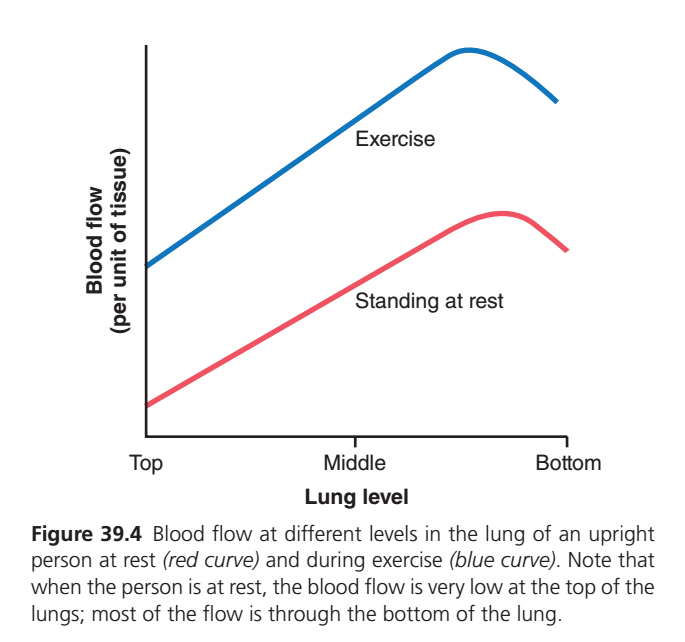
Figure 39.4: Blood Flow at Different Levels of the Lung (Guyton Physiology 15th Edition)
🎯 One-Line Core Concept
In an upright person, blood flow is lowest at the top (apex) of the lungs and highest at the bottom (base) because gravity pulls blood downward. During exercise, blood flow increases throughout the lungs, especially at the top, making blood distribution more uniform.
🧠 First Understand the Big Picture
Imagine holding a water bottle vertically.
Where does most of the water collect?
✅ At the bottom because of gravity.
Blood behaves exactly the same inside the lungs.
When you stand upright:
- Less blood reaches the top (apex).
- More blood reaches the bottom (base).
This is the main idea of the graph.
Step 1: Understanding the Axes
Y-axis (Vertical)
Blood Flow (per unit of tissue)
This tells us:
How much blood reaches each part of the lung.
X-axis (Horizontal)
Lung Level
This represents different heights of the lung.
Top (Apex)
      ↓
Middle
      ↓
Bottom (Base)
It is not time.
It simply shows different levels of the lung from top to bottom.
There Are Two Curves
🔴 Red Curve
Standing at Rest
Shows blood flow while a person is:
- Standing
- Relaxed
- Not exercising
🔵 Blue Curve
Exercise
Shows blood flow while the person is exercising.
PART 1 — Red Curve (Standing at Rest)
Let’s follow the red line from left to right.
Top of the Lung (Apex)
Notice the red curve starts very low.
What does this mean?
Very little blood reaches the top of the lungs.
Why?
Because gravity pulls blood downward.
The blood has to move upward against gravity.
Therefore,
only a small amount reaches the apex.
Important Concept
The apex is well ventilated (air reaches it easily),
but it is poorly perfused (receives less blood).
Middle of the Lung
The red curve gradually rises.
Why?
As we move downward,
gravity becomes less of a problem.
More blood reaches these lung regions.
Therefore,
blood flow increases.
Bottom of the Lung (Base)
Here the red curve reaches its highest point.
Why?
Gravity pulls blood toward the lower lung.
The pulmonary vessels here contain more blood.
Therefore,
the base receives the greatest blood flow.
Easy Memory
Top Lung
↓
Least Blood

Middle Lung
↓
Moderate Blood

Bottom Lung
↓
Maximum Blood
Why Does the Red Curve Slightly Fall at the End?
Notice the curve bends downward slightly near the extreme base.
This represents that the very lowest part of the lung does not continue to receive endlessly increasing blood flow.
Why?
At the extreme base:
- Lung tissue becomes more compressed by the weight of the lung itself.
- Pulmonary vessels can become slightly compressed.
- Therefore, blood flow reaches a maximum and then decreases slightly.
The important clinical message remains:
The base of the lung still receives much more blood than the apex.
PART 2 — Blue Curve (Exercise)
Now look at the blue curve.
It starts much higher than the red curve.
Why Is It Higher?
During exercise:
The heart pumps much more blood.
Cardiac output increases.
More blood enters the pulmonary circulation.
Therefore,
blood flow increases throughout the lungs.
Top of the Lung During Exercise
Notice something important.
The blue curve is much higher than the red curve.
Why?
During exercise:
Previously closed or partially closed pulmonary capillaries open.
This is called:
Capillary Recruitment
Also,
already open vessels become wider.
This is called:
Capillary Distension
Together,
these changes greatly increase blood flow at the apex.
Middle of the Lung
Blood flow continues to increase.
Now almost the entire lung receives abundant blood.
Bottom of the Lung
The base still receives the highest blood flow.
However,
the difference between the top and bottom becomes much smaller.
Why?
Exercise distributes blood more evenly throughout the lungs.
Slight Fall at the End
The blue curve also bends slightly downward near the extreme base.
For the same reasons as at rest:
- Mild vessel compression
- Mechanical effects of lung tissue
Despite this,
blood flow remains much higher than during rest.
Why Does Exercise Increase Blood Flow?
During exercise:
- Muscles need more oxygen.
- Cardiac output increases.
- More blood enters the pulmonary arteries.
- Pulmonary vessels recruit and distend.
- Pulmonary vascular resistance falls.
- More blood reaches every part of the lung.
This improves oxygen uptake.
Why Doesn’t Blood Stay Mostly at the Bottom During Exercise?
Normally,
gravity causes blood to collect at the base.
During exercise,
the increased pressure inside pulmonary arteries opens many capillaries in the upper lung.
As a result,
blood distribution becomes more uniform.
Rest vs Exercise
| Feature | Standing at Rest | During Exercise |
|---|---|---|
| Cardiac output | Normal | Increased |
| Blood flow at apex | Very low | Markedly increased |
| Blood flow at base | Highest | Still highest |
| Blood distribution | Uneven | More uniform |
| Pulmonary capillaries | Many closed | Recruited and distended |
Why Is This Physiologically Important?
If only the base received blood during exercise,
the lungs would not oxygenate the greatly increased cardiac output efficiently.
By opening capillaries throughout the lungs,
the body uses a much larger surface area for gas exchange.
This helps maintain excellent oxygenation even during heavy exercise.
Easy Story to Remember
Imagine a three-floor building:
- Top floor = Apex
- Middle floor = Middle lung
- Ground floor = Base
At Rest
A delivery truck (blood) has limited packages.
Most deliveries are made to the ground floor because it is easiest to reach.
Very few reach the top floor.
During Exercise
Now many more delivery trucks arrive.
They can deliver packages to all floors, including the top.
So every floor receives better supplies.
This is exactly how pulmonary blood flow changes.
Easy Flowchart
Standing Upright
        ↓
Gravity pulls blood downward
        ↓
Least blood at Apex
        ↓
Most blood at Base
During Exercise
Exercise
        ↓
↑ Cardiac Output
        ↓
Capillary Recruitment + Distension
        ↓
↑ Blood Flow Throughout Lung
        ↓
More Uniform Perfusion
        ↓
Better Gas Exchange
📚 High-Yield MBBS Points
- Gravity is the main reason blood flow is lowest at the apex and highest at the base of the lungs in an upright person.
- At rest (red curve): Blood flow progressively increases from the apex to the base.
- During exercise (blue curve): Blood flow increases throughout the lungs because of increased cardiac output, capillary recruitment, and capillary distension.
- The difference in blood flow between the apex and base becomes smaller during exercise, resulting in a more even distribution of perfusion.
- Clinical significance: More uniform pulmonary blood flow during exercise improves ventilation–perfusion (V/Q) matching and enhances oxygen uptake despite the increased demand.
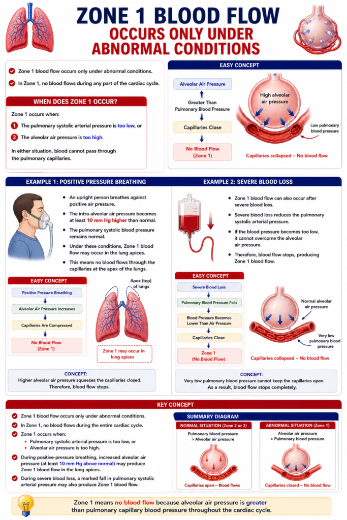
Zones 1, 2, and 3 of Pulmonary Blood Flow
- The pulmonary capillaries are located in the walls of the alveoli.
- These capillaries are pushed outward by the blood pressure inside them.
- At the same time, they are compressed by the alveolar air pressure from outside.
- If the alveolar air pressure becomes greater than the capillary blood pressure, the capillaries collapse (close).
- When the capillaries close, blood flow stops.
- Depending on normal or disease conditions, the lungs may have different zones of pulmonary blood flow.
- These zones are divided into three patterns.
Zone 1
- No blood flow occurs during any part of the cardiac cycle.
- This is because the alveolar capillary pressure never becomes higher than the alveolar air pressure.
- Therefore, the capillaries remain closed throughout the cardiac cycle.
Easy Concept
Alveolar Air Pressure
        ▲
        │
Greater Than
Capillary Blood Pressure
        │
        ▼
Capillaries Close
        │
        ▼
No Blood Flow
(Zone 1)
Concept:
- Air pressure is always greater than blood pressure.
- Therefore, blood cannot pass through the capillaries.
Zone 2
- Blood flows only during systole.
- During systole, the pulmonary arterial pressure becomes greater than the alveolar air pressure.
- During diastole, the pulmonary arterial pressure becomes lower than the alveolar air pressure.
- Therefore, blood flow is intermittent.
Easy Concept
SYSTOLE
Blood Pressure > Air Pressure
        │
        ▼
Blood Flows

DIASOLE
Blood Pressure < Air Pressure
        │
        ▼
No Blood Flow
Concept:
- Flow during systole
- No flow during diastole
- Therefore, blood flow is intermittent.
Zone 3
- Blood flows continuously throughout the cardiac cycle.
- During both systole and diastole, the alveolar capillary pressure remains greater than the alveolar air pressure.
- Therefore, the capillaries remain open continuously.
Easy Concept
SYSTOLE
Blood Pressure > Air Pressure
        │
        ▼
Blood Flows

DIASOLE
Blood Pressure > Air Pressure
        │
        ▼
Blood Continues to Flow
Concept:
- Blood pressure is always higher than air pressure.
- Therefore, continuous blood flow occurs.
Normal Distribution of Pulmonary Blood Flow
- In normal lungs, only Zone 2 and Zone 3 are present.
- Zone 2 is found in the apex (top) of the lungs.
- Zone 3 is present in the lower parts of the lungs.
Zone 2 in the Apex of the Lung
- In a standing person, the pulmonary arterial pressure at the lung apex is about 15 mm Hg lower than at the level of the heart.
- Therefore, the apical systolic pressure is about 10 mm Hg.
Step-by-Step Understanding
Step 1: Pressure at Heart Level
Pulmonary arterial systolic pressure = 25 mm Hg
↓
Step 2: Pressure Lost Due to Hydrostatic Effect
Hydrostatic pressure difference = 15 mm Hg
↓
Step 3: Calculate the Pressure at the Apex
Apical Systolic Pressure = 25 − 15
= 10 mm Hg
Final Answer
Apical systolic pressure = 10 mm Hg
Easy Concept
Heart Level
25 mm Hg
      │
Minus 15 mm Hg
(Hydrostatic Pressure)
      │
      ▼
Lung Apex
10 mm Hg
Concept:
- Blood loses 15 mm Hg while moving upward.
- Therefore, the pressure at the lung apex becomes 10 mm Hg.
Blood Flow During Systole
- The 10 mm Hg systolic pressure is greater than the alveolar air pressure (0 mm Hg).
- Therefore, blood flows during systole.
Blood Flow During Diastole
- The diastolic pulmonary arterial pressure at heart level is 8 mm Hg.
- This pressure cannot overcome the 15 mm Hg hydrostatic pressure difference needed to reach the lung apex.
- Therefore, no blood flows during diastole.
Step-by-Step Understanding
During Systole
10 mm Hg > 0 mm Hg
↓
Blood flows
During Diastole
8 mm Hg < 15 mm Hg hydrostatic pressure difference
↓
Blood cannot reach the apex
↓
No blood flow
Why Is This Called Zone 2?
- Blood flows only during systole.
- Blood stops flowing during diastole.
- Therefore, Zone 2 has intermittent blood flow.
Location of Zone 2
- Zone 2 begins about 10 cm above the level of the heart.
- It extends from that level to the apex of the lungs.
Zone 3 in the Lower Lung
- In the lower regions of the lungs, the pulmonary arterial pressure remains greater than the alveolar air pressure during both systole and diastole.
- Therefore, continuous blood flow occurs.
- This is called Zone 3 blood flow.
Easy Concept
Lower Lung

Systole
Blood Pressure > Air Pressure
        │
        ▼
Blood Flows

Diastole
Blood Pressure > Air Pressure
        │
        ▼
Blood Continues to Flow

Continuous Flow
(Zone 3)
Lying Down (Supine Position)
- When a person lies down, no part of the lung is more than a few centimeters above the level of the heart.
- Therefore, the hydrostatic pressure difference becomes very small.
Easy Concept
Standing
Large Height Difference
        │
        ▼
Zone 2 + Zone 3

Lying Down
Very Small Height Difference
        │
        ▼
Mostly Continuous Blood Flow
Concept:
- In the standing position, gravity creates different blood flow zones.
- In the lying position, gravity has much less effect because the lungs are nearly at the same level as the heart.
Key Concept
- Pulmonary capillaries remain open only when capillary blood pressure is greater than alveolar air pressure.
- Zone 1: No blood flow because alveolar air pressure is always greater than capillary pressure.
- Zone 2:Intermittent blood flow because:
- Systolic pressure > alveolar air pressure
- Diastolic pressure < alveolar air pressure
- Zone 3: Continuous blood flow because capillary pressure remains greater than alveolar air pressure throughout the cardiac cycle.
- In normal lungs:
- Zone 2 is found in the apex.
- Zone 3 is found in the lower lung.
- Apical systolic pressure = 25 − 15 = 10 mm Hg.
- Zone 2 begins about 10 cm above the level of the heart and extends to the top of the lungs.
- In the lower lungs, pulmonary arterial pressure remains greater than alveolar air pressure during both systole and diastole, producing continuous (Zone 3) blood flow.
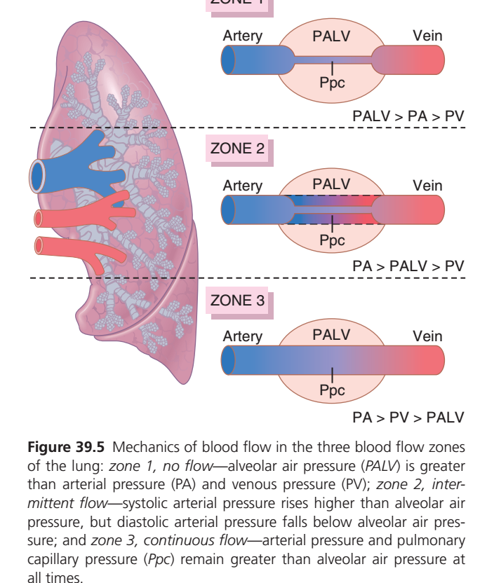
Figure 39.5: Blood Flow Zones of the Lung (Guyton Physiology 15th Edition)
🎯 One-Line Core Concept
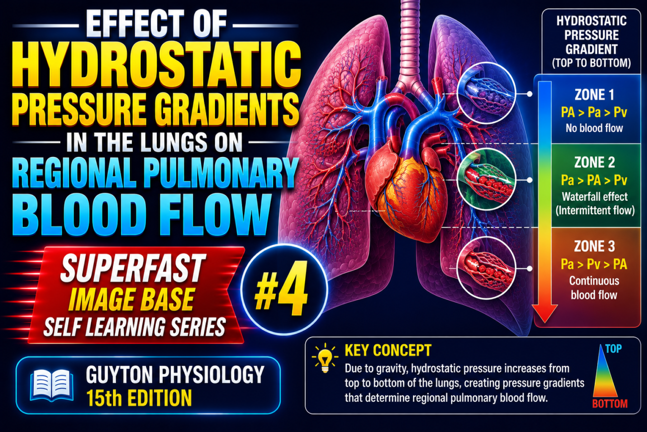
Gravity causes blood pressure to be different at the top, middle, and bottom of the lungs. Depending on the relationship between alveolar air pressure and blood pressure, the lung is divided into three zones:
- Zone 1 = No blood flow
- Zone 2 = Intermittent blood flow
- Zone 3 = Continuous blood flow
This is one of the most important MBBS physiology concepts because it explains how gravity affects pulmonary blood flow.
🧠 First Understand the Big Picture
Imagine you are holding a vertical water pipe.
At the top of the pipe, water pressure is low.
At the bottom, water pressure is high because gravity pulls water downward.
Blood behaves exactly the same inside the lungs.
Therefore:
- Top of lung → Lowest blood pressure
- Middle of lung → Moderate blood pressure
- Bottom of lung → Highest blood pressure
Meanwhile,
Alveolar air pressure (PALV) remains almost the same throughout the lung.
This difference creates the three lung zones.
Before Understanding the Zones, Learn These Four Pressures
1. PA = Pulmonary Arterial Pressure
This is the pressure of blood entering the pulmonary capillary.
Think of it as the entry pressure.
2. PV = Pulmonary Venous Pressure
This is the pressure of blood leaving the pulmonary capillary.
Think of it as the exit pressure.
3. PALV = Alveolar Air Pressure
This is the pressure of the air inside the alveolus.
It pushes from outside on the capillaries.
Unlike blood pressure,
PALV remains almost constant throughout the lung.
4. Ppc = Pulmonary Capillary Pressure
This is the pressure inside the pulmonary capillary.
It lies between arterial and venous pressure.
Why Does Blood Flow Stop?
Imagine squeezing a garden hose.
If the pressure outside the hose becomes greater than the water pressure inside,
the hose collapses.
Blood can no longer flow.
Exactly the same thing happens in the lung.
If
PALV > Blood Pressure
↓
Capillary collapses
↓
Blood flow stops.
THE LUNG IS DIVIDED INTO THREE ZONES
Top
↓
ZONE 1

Middle
↓
ZONE 2

Bottom
↓
ZONE 3
🔴 ZONE 1 (Top of Lung)
Look at the upper part of the figure.
The relationship shown is
PALV > PA > PV
Let’s understand this carefully.
What does this mean?
The pressure inside the alveolus is greater than both arterial and venous blood pressure.
Alveolar Air Pressure = 15

Arterial Pressure = 10

Venous Pressure = 5
Since air pressure is highest,
the capillary is squeezed closed.
Like stepping on a hose,
blood cannot pass.
Result
🚫 No Blood Flow
Why?
Air pressure compresses the pulmonary capillary.
Blood cannot enter.
Is Zone 1 Normally Present?
No.
In a healthy person,
Zone 1 is usually absent or extremely small.
It appears when:
- Severe blood loss
- Shock
- Positive-pressure ventilation
- Mechanical ventilation with high airway pressures
Easy Memory
Zone 1 = Air Wins
Air pressure is highest.
Blood loses.
No blood flows.
🟡 ZONE 2 (Middle of Lung)
Relationship shown:
PA > PALV > PV
This is the most confusing zone for students.
Let’s simplify it.
During Heart Contraction (Systole)
Arterial pressure rises.
Suppose
PA = 25

PALV = 15

PV = 8
Now
PA > PALV
Blood can enter the capillary.
Blood flows.
During Heart Relaxation (Diastole)
Arterial pressure falls.
Suppose
PA = 10

PALV = 15

PV = 5
Now
PALV > PA
The capillary is compressed.
Blood flow stops.
Result
Blood flows only during systole.
Stops during diastole.
Therefore,
Flow is
Intermittent
Easy Analogy
Imagine a railway crossing.
The gate opens only when the train arrives.
Otherwise it stays closed.
Similarly,
Blood flows only when arterial pressure becomes higher than alveolar pressure.
Easy Memory
Zone 2 = Sometimes Flow
Heart beats
↓
Blood flows
Heart relaxes
↓
Blood stops
🟢 ZONE 3 (Bottom of Lung)
Relationship shown:
PA > PV > PALV
This is the normal situation.
Suppose
PA = 25

PV = 12

PALV = 8
Now,
Both arterial and venous pressures are higher than alveolar pressure.
The capillary remains open all the time.
Nothing compresses it.
Blood flows continuously.
Result
✅ Continuous Blood Flow
Why?
Gravity increases blood pressure toward the base of the lung.
Therefore,
Both arterial and venous pressures remain higher than alveolar pressure throughout the cardiac cycle.
Easy Memory
Zone 3 = Blood Wins
Blood pressure is always stronger than air pressure.
Flow never stops.
What Happens From Top to Bottom?
| Lung Region | Blood Pressure | Blood Flow |
|---|---|---|
| Apex | Lowest | Very little |
| Middle | Moderate | Intermittent |
| Base | Highest | Continuous |
Why Does Gravity Cause This?
Blood has weight.
Gravity pulls blood downward.
Therefore:
At the top,
blood pressure is very low.
At the bottom,
blood pressure becomes much higher.
Alveolar air pressure hardly changes.
That is why the relationship between these pressures changes from top to bottom.
Easy Flowchart
Standing Person
        ↓
Gravity
        ↓
Blood pressure decreases toward apex
        ↓
Blood pressure increases toward base
        ↓
Three Lung Zones
Zone Summary
🔴 Zone 1
PALV > PA > PV↓
Capillary collapsed
↓
🚫 No blood flow
🟡 Zone 2
PA > PALV > PV↓
Blood flows only when arterial pressure exceeds alveolar pressure
↓
⚠️ Intermittent blood flow
🟢 Zone 3
PA > PV > PALV↓
Capillary always open
↓
✅ Continuous blood flow
Memory Trick
Think of Air vs Blood.
Zone 1
Air is strongest.
🚫 No flow.
Zone 2
Air and blood take turns winning.
⚠️ Intermittent flow.
Zone 3
Blood is strongest.
✅ Continuous flow.Why Is This Clinically Important?
Zone 1
Seen in:
- Severe hemorrhage
- Shock
- Positive-pressure ventilation
Blood pressure falls or alveolar pressure rises enough to collapse capillaries.
Zone 2
Represents the transition region where flow depends on the cardiac cycle.
Zone 3
This is the largest and most important zone in healthy lungs.
Most pulmonary blood flow occurs here, making it the major site for efficient gas exchange.
🎓 High-Yield MBBS Points
- Gravity causes pulmonary arterial and venous pressures to increase from the apex to the base of the lungs, while alveolar air pressure (PALV) remains nearly constant.
- Zone 1 (PALV > PA > PV): Alveolar pressure compresses pulmonary capillaries, causing no blood flow. It is usually absent in healthy individuals but may appear in shock or during positive-pressure ventilation.
- Zone 2 (PA > PALV > PV): Blood flows intermittently, mainly during systole when pulmonary arterial pressure exceeds alveolar pressure. This is known as the “waterfall effect” because flow depends on the difference between PA and PALV, not on venous pressure.
- Zone 3 (PA > PV > PALV): Both arterial and venous pressures remain higher than alveolar pressure, so capillaries stay open and blood flows continuously. This is the normal condition in the lower lungs and is where most pulmonary perfusion occurs.
- Easy memory: Zone 1 = No Flow, Zone 2 = Intermittent Flow, Zone 3 = Continuous Flow.
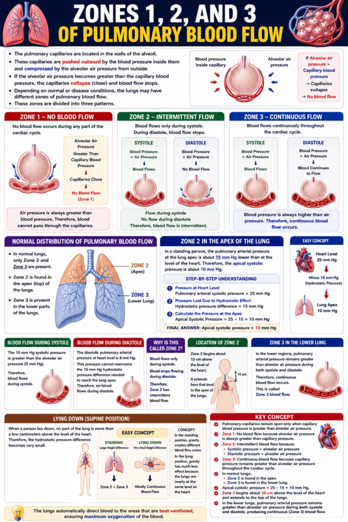
Zone 1 Blood Flow Occurs Only Under Abnormal Conditions
- Zone 1 blood flow occurs only under abnormal conditions.
- In Zone 1, no blood flows during any part of the cardiac cycle.
When Does Zone 1 Occur?
- Zone 1 occurs when:
- The pulmonary systolic arterial pressure is too low, or
- The alveolar air pressure is too high.
- In either situation, blood cannot pass through the pulmonary capillaries.
Easy Concept
Think of the alveolar air pressure pressing on the capillaries.
Alveolar Air Pressure
        ▲
        │
Greater Than
Pulmonary Blood Pressure
        │
        ▼
Capillaries Close
        │
        ▼
No Blood Flow
(Zone 1)
Concept:
- If air pressure becomes greater than blood pressure, the capillaries collapse.
- Therefore, blood cannot flow.
Example 1: Positive Pressure Breathing
- An upright person breathes against positive air pressure.
- The intra-alveolar air pressure becomes at least 10 mm Hg higher than normal.
- The pulmonary systolic blood pressure remains normal.
- Under these conditions, Zone 1 blood flow may occur in the lung apices.
- This means no blood flows through the capillaries at the apex of the lungs.
Easy Concept
Positive Pressure Breathing
        │
        ▼
Alveolar Air Pressure Increases
        │
        ▼
Capillaries Are Compressed
        │
        ▼
No Blood Flow
(Zone 1)
Concept:
- Higher alveolar air pressure squeezes the capillaries closed.
- Therefore, blood flow stops.
Example 2: Severe Blood Loss
- Zone 1 blood flow can also occur after severe blood loss.
- Severe blood loss reduces the pulmonary systolic arterial pressure.
- If the blood pressure becomes too low, it cannot overcome the alveolar air pressure.
- Therefore, blood flow stops, producing Zone 1 blood flow.
Easy Concept
Severe Blood Loss
        │
        ▼
Pulmonary Blood Pressure Falls
        │
        ▼
Blood Pressure Becomes
Lower Than Air Pressure
        │
        ▼
Capillaries Close
        │
        ▼
Zone 1
(No Blood Flow)
Concept:
- Very low pulmonary blood pressure cannot keep the capillaries open.
- As a result, blood flow stops completely.
Key Concept
- Zone 1 blood flow occurs only under abnormal conditions.
- In Zone 1, no blood flows during the entire cardiac cycle.
- Zone 1 occurs when:
- Pulmonary systolic arterial pressure is too low, or
- Alveolar air pressure is too high.
- During positive-pressure breathing, increased alveolar air pressure (at least 10 mm Hg above normal) may produce Zone 1 blood flow in the lung apices.
- During severe blood loss, a marked fall in pulmonary systolic arterial pressure may also produce Zone 1 blood flow.
Exercise Normally Increases Blood Flow Through All Parts of the Lungs
Figure: Fig. 39.4
- Fig. 39.4 shows that blood flow increases in all parts of the lungs during exercise.
- During exercise, the pulmonary vascular pressures increase.
Effect of Exercise on Pulmonary Blood Flow
- The increase in pulmonary vascular pressure changes the pattern of blood flow in the lung apices.
- During rest, the lung apices normally have Zone 2 blood flow.
- During exercise, the lung apices change from Zone 2 to Zone 3 blood flow.
Easy Concept
At Rest
Rest
      │
      ▼
Pulmonary Vascular Pressure Lower
      │
      ▼
Lung Apex
Zone 2
(Intermittent Blood Flow)
Concept:
- At rest, the pressure is not high enough to maintain continuous blood flow at the lung apex.
- Therefore, the apex has Zone 2 (intermittent) blood flow.
During Exercise
Exercise
      │
      ▼
Pulmonary Vascular Pressure Increases
      │
      ▼
More Blood Reaches Lung Apex
      │
      ▼
Zone 2
        ↓
Zone 3
(Continuous Blood Flow)
Concept:
- During exercise, pulmonary vascular pressure rises.
- The higher pressure keeps the pulmonary capillaries open throughout the cardiac cycle.
- Therefore, the lung apex changes from Zone 2 to Zone 3 blood flow.
Why Does Blood Flow Increase During Exercise?
Exercise
      │
      ▼
Pulmonary Vascular Pressure Increases
      │
      ▼
Capillaries Stay Open
Throughout the Cardiac Cycle
      │
      ▼
Continuous Blood Flow
(Zone 3)
      │
      ▼
Blood Flow Increases
Throughout the Lungs
Concept:
- Higher pulmonary vascular pressure allows continuous blood flow, even in the upper parts of the lungs.
- As a result, all regions of the lungs receive more blood during exercise.
Key Concept
- Fig. 39.4 shows that exercise increases blood flow throughout the lungs.
- During exercise, pulmonary vascular pressure rises.
- The increased pressure converts the lung apices from Zone 2 (intermittent blood flow) to Zone 3 (continuous blood flow).
- This results in greater blood flow through all parts of the lungs.
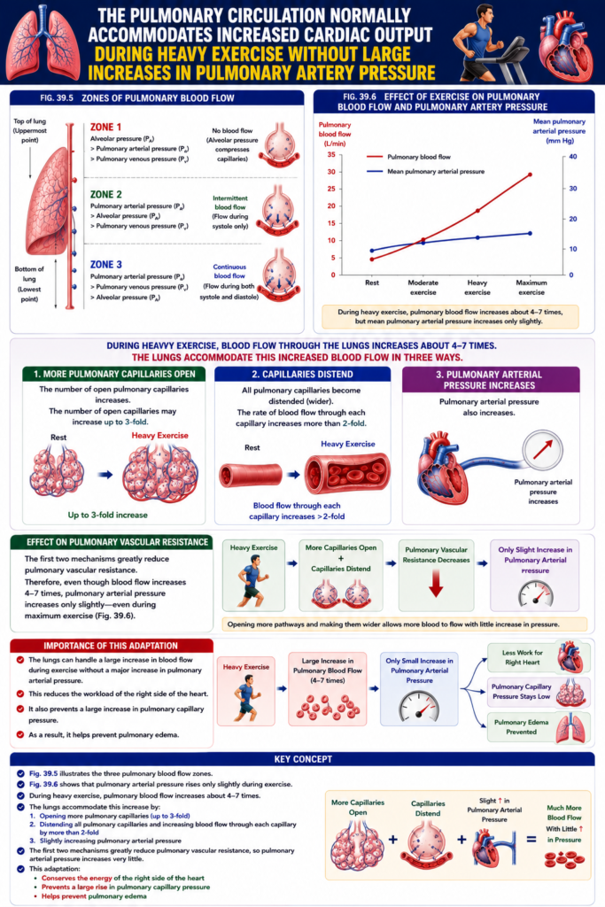
The Pulmonary Circulation Normally Accommodates Increased Cardiac Output During Heavy Exercise Without Large Increases in Pulmonary Artery Pressure
Figure: Fig. 39.5 and Fig. 39.6
- During heavy exercise, blood flow through the lungs increases about 4–7 times.
- The lungs accommodate this increased blood flow in three ways.
Method 1: More Pulmonary Capillaries Open
- The number of open pulmonary capillaries increases.
- The number of open capillaries may increase up to 3-fold.
Method 2: Capillaries Distend
- All pulmonary capillaries become distended (wider).
- The rate of blood flow through each capillary increases more than 2-fold.
Method 3: Pulmonary Arterial Pressure Increases
- Pulmonary arterial pressure also increases.
Easy Concept
Heavy Exercise
        │
        ▼
Blood Flow Through Lungs
Increases 4–7 Times
        │
        ▼
Three Adaptations
1. More Capillaries Open
│
▼
Up to 3-Fold Increase2. Capillaries Distend
│
▼
Blood Flow Through Each
Capillary Increases
(More Than 2-Fold)3. Pulmonary Arterial
Pressure IncreasesConcept:
- The lungs do not rely on only one mechanism.
- They open more capillaries, widen existing capillaries, and slightly increase pulmonary arterial pressure to handle the increased blood flow.
Effect on Pulmonary Vascular Resistance
- Normally, the first two mechanisms greatly reduce pulmonary vascular resistance.
- Because resistance decreases so much, the pulmonary arterial pressure increases only slightly, even during maximum exercise.
- This effect is shown in Fig. 39.6.
Easy Concept
Heavy Exercise
        │
        ▼
More Capillaries Open
+
Capillaries Distend
        │
        ▼
Pulmonary Vascular
Resistance Decreases
        │
        ▼
Only Slight Increase in
Pulmonary Arterial Pressure
Concept:
- Opening more pathways and making them wider allows more blood to flow with little increase in pressure.
Importance of This Adaptation
- The lungs can handle a large increase in blood flow during exercise without a major increase in pulmonary arterial pressure.
- This reduces the workload of the right side of the heart.
- It also prevents a large increase in pulmonary capillary pressure.
- As a result, it helps prevent pulmonary edema.
Easy Concept
Heavy Exercise
        │
        ▼
Large Increase in
Pulmonary Blood Flow
        │
        ▼
Only Small Increase in
Pulmonary Arterial Pressure
        │
        ├──────────────┐
        ▼              ▼
Less Work for     Pulmonary Capillary
Right Heart       Pressure Stays Low
                         │
                         ▼
              Pulmonary Edema Prevented
Concept:
- The pulmonary circulation is highly adaptable.
- It allows much more blood flow while keeping pressure almost unchanged.
- This protects both the right heart and the lungs.
Key Concept
- Fig. 39.5 illustrates the three pulmonary blood flow zones.
- Fig. 39.6 shows that pulmonary arterial pressure rises only slightly during exercise.
- During heavy exercise, pulmonary blood flow increases about 4–7 times.
- The lungs accommodate this increase by:
- Opening more pulmonary capillaries (up to 3-fold)
- Distending all pulmonary capillaries and increasing blood flow through each capillary by more than 2-fold
- Slightly increasing pulmonary arterial pressure
- The first two mechanisms greatly reduce pulmonary vascular resistance, so pulmonary arterial pressure increases very little.
- This adaptation:
- Conserves the energy of the right side of the heart
- Prevents a large rise in pulmonary capillary pressure
- Helps prevent pulmonary edema
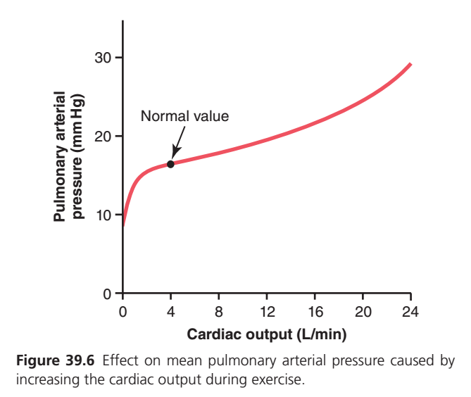
Understanding Fig. 39.6 (Guyton Physiology 15th Ed.)
Effect of Increasing Cardiac Output on Mean Pulmonary Arterial Pressure During Exercise
Figure: Fig. 39.6
Step 1: Understand the Axes
Horizontal (X-axis)
Cardiac Output (L/min)
- Shows how much blood the heart pumps every minute.
- Units = Liters/minute (L/min)
Left -----------------------------> Right

Low Cardiac Output          High Cardiac Output

0      4      8      12      16      20      24
Concept
➡️ Moving to the right means
Heart pumps more blood every minute.
Vertical (Y-axis)
Pulmonary Arterial Pressure (mm Hg)
- Shows the average pressure inside the pulmonary artery.
Top
↑
High Pulmonary Pressure
30 mm Hg
25
20
15
10
0
↓
Bottom
Low Pulmonary Pressure
Concept
➡️ Moving upward means
Pulmonary artery pressure is increasing.
Step 2: Normal Value
The black dot represents the normal resting condition.
Cardiac Output ≈ 5 L/min

↓

Mean Pulmonary Arterial Pressure
≈15 mm Hg
This is the normal value.
Easy Memory
REST

Heart pumps
≈5 L/min

↓

Pulmonary artery pressure

≈15 mm Hg
Step 3: First Part of the Curve
Look at the left side.
Pressure
30 |
25 |
20 |
15 |____
10 |
   +----------------
      0   2   4
Notice
The curve rises very quickly at first.
Why?
When blood first enters the lungs,
many pulmonary capillaries are closed.
As cardiac output increases,
more pulmonary capillaries open.
Guyton calls this
Capillary Recruitment
Easy Concept
Small Blood Flow

↓

Few Capillaries Open

↓

Increase Blood Flow

↓

More Capillaries Open

↓

Pressure rises only slightly
after the initial increase
Step 4: Middle of the Curve
Between about
5–16 L/min
look carefully.
The curve becomes almost flat.
Pressure

20 |
19 |
18 |
17 |
16 |---------
15 |
What does this mean?
Even though
cardiac output is increasing greatly,
pulmonary arterial pressure increases only a little.
Why?
Because the lungs do two amazing things.
Mechanism 1
More capillaries open.
↓
Mechanism 2
Already open capillaries become wider.
↓
Resistance falls.
↓
Pressure hardly rises.
Easy Flow
Exercise

↓

Cardiac Output ↑

↓

More Capillaries Open

+

Capillaries Stretch

↓

Resistance ↓

↓

Pressure stays almost normal
This is the main concept of this graph.
Step 5: Right Side of the Curve
Now look at the far right.
Around
20–24 L/min.
The curve begins to rise more.
Pressure

30 |
28 |
26 |
24 |
22 |
20 |
Why?
Now almost
✔ all capillaries are already open
✔ all capillaries are already stretched
The lungs cannot enlarge much more.
Therefore,
extra blood causes
pressure to increase faster.asy Concept
Maximum Exercise

↓

Almost Every Capillary
Already Open

↓

Cannot Recruit More

↓

Pressure Begins
to Rise Faster
Step 6: Whole Story of the Graph
Rest
↓

Cardiac Output
≈5 L/min

↓

Pressure
≈15 mm Hg

────────────────────────

Exercise Starts

↓

More Blood

↓

More Capillaries Open

↓

Capillaries Stretch

↓

Resistance Falls

↓

Pressure Hardly Increases

────────────────────────

Very Heavy Exercise

↓

Almost Every Capillary
Already Open

↓

No More Reserve

↓

Pressure Starts Rising Faster
Clinical Importance (Very High Yield)
Why doesn’t pulmonary artery pressure rise much during exercise?
Because the lungs:
- Open more pulmonary capillaries (recruitment)
- Stretch existing capillaries (distension)
↓
Pulmonary vascular resistance decreases.
↓
Large increase in blood flow occurs with only a small increase in pressure.
What would happen if this mechanism did NOT exist?
Exercise

↓

Large Increase
in Pulmonary Pressure

↓

Pulmonary Capillary Pressure ↑

↓

Fluid Leaks into Alveoli

↓

Pulmonary Edema

↓

Poor Gas Exchange
Super Memory Trick (Guyton Favorite)
Think of the pulmonary circulation as a highway.
Rest

10 Roads Open

↓

Exercise

20 Roads Open

↓

Cars (Blood)

Spread Out

↓

Traffic Jam
(Pressure)

Does NOT Increase Much
Only when every road is already open does adding more cars cause a traffic jam (pressure increase).
Key Concept
- Fig. 39.6 shows the relationship between cardiac output and mean pulmonary arterial pressure.
- At rest, cardiac output ≈ 5 L/min and mean pulmonary arterial pressure ≈ 15 mm Hg.
- During exercise, cardiac output increases markedly, but pulmonary arterial pressure rises only slightly because:
- More pulmonary capillaries open (capillary recruitment).
- Existing capillaries distend (capillary distension).
- These changes reduce pulmonary vascular resistance, allowing 4–7 times more blood flow with only a small increase in pressure.
- During very heavy exercise, most capillaries are already open and fully distended, so pulmonary arterial pressure begins to rise more rapidly.
- This adaptation reduces the workload of the right ventricle and helps prevent pulmonary edema.
Function of Pulmonary Circulation When Left Atrial Pressure Rises as a Result of Left-Sided Heart Failure
- In a healthy person, the left atrial pressure almost never rises above +6 mm Hg, even during very strenuous exercise.
- These small increases in left atrial pressure have almost no effect on pulmonary circulation.
- This is because:
- The pulmonary venules expand.
- More pulmonary capillaries open.
- As a result, blood continues to flow easily from the pulmonary arteries.
Left-Sided Heart Failure
- When the left side of the heart fails, blood begins to accumulate (dam up) in the left atrium.
- As a result, the left atrial pressure increases.
- The normal left atrial pressure is 1–5 mm Hg.
- In left-sided heart failure, it may rise to 40–50 mm Hg.
Easy Concept
Healthy Left Heart
        │
        ▼
Left Atrial Pressure
1–5 mm Hg
        │
        ▼
Blood Flows Easily
Through the Lungs
Left-Sided Heart Failure
│
▼
Blood Backs Up
in Left Atrium
│
▼
Left Atrial Pressure
Rises
(40–50 mm Hg)Concept:
- A healthy left heart allows blood to leave the lungs easily.
- In left-sided heart failure, blood cannot leave the left atrium efficiently, so it backs up into the lungs.
Effect of Mild Increase in Left Atrial Pressure
- When the left atrial pressure rises up to about 7 mm Hg, it has little effect on pulmonary circulation.
Easy Concept
Left Atrial Pressure
≤ 7 mm Hg
        │
        ▼
Pulmonary Venules Expand
+
More Capillaries Open
        │
        ▼
Pulmonary Blood Flow
Remains Normal
Concept:
- Up to 7 mm Hg, the lungs adjust by opening more capillaries.
- Therefore, blood continues to flow normally.
Effect of Left Atrial Pressure Greater Than 7–8 mm Hg
- When the left atrial pressure rises above 7–8 mm Hg, pulmonary arterial pressure also increases almost equally.
- This increases the workload on the right side of the heart.
- Pulmonary capillary pressure also increases almost equally.
Easy Concept
Left Atrial Pressure
>7–8 mm Hg
        │
        ▼
Pulmonary Arterial
Pressure Increases
        │
        ▼
Pulmonary Capillary
Pressure Increases
        │
        ▼
Right Heart Works Harder
Concept:
- After 7–8 mm Hg, pressure is transmitted backward into the pulmonary circulation.
- This raises both pulmonary arterial pressure and pulmonary capillary pressure.
Development of Pulmonary Edema
- When the left atrial pressure rises above 30 mm Hg, the pulmonary capillary pressure also rises to a similar level.
- At this stage, pulmonary edema is likely to develop.
Easy Concept
Left Atrial Pressure
>30 mm Hg
        │
        ▼
Pulmonary Capillary
Pressure Increases
        │
        ▼
Pulmonary Edema
Likely to Develop
Concept:
- Very high left atrial pressure causes very high pulmonary capillary pressure.
- This makes pulmonary edema likely.
Key Concept
- In a healthy person, left atrial pressure rarely exceeds +6 mm Hg, even during strenuous exercise.
- Small increases in left atrial pressure have little effect because pulmonary venules expand and more capillaries open.
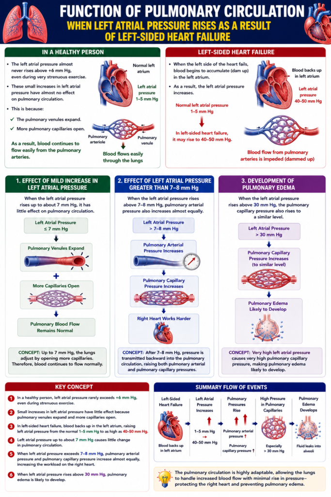
- In left-sided heart failure, blood backs up in the left atrium, raising left atrial pressure from the normal 1–5 mm Hg to as high as 40–50 mm Hg.
- Left atrial pressure up to about 7 mm Hg causes little change in pulmonary circulation.
- When left atrial pressure exceeds 7–8 mm Hg, pulmonary arterial pressure and pulmonary capillary pressure increase almost equally, increasing the workload on the right heart.
- When left atrial pressure rises above 30 mm Hg, pulmonary edema is likely to develop.