


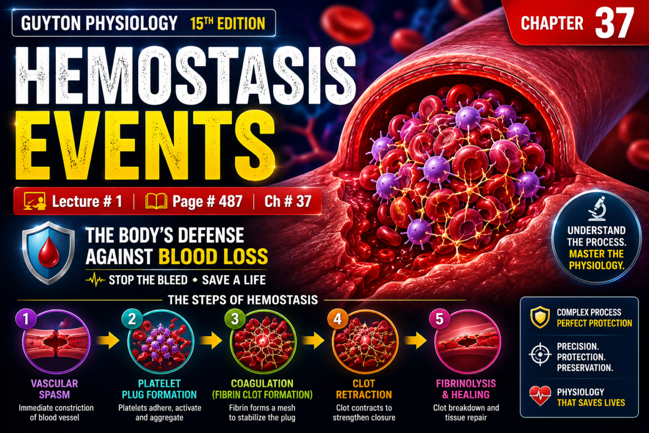
- Hemostasis is the process that prevents blood loss.
- When a blood vessel is cut (severed) or damaged (ruptured), the body stops bleeding through several mechanisms.
- Mechanism 1: The blood vessel constricts (becomes narrow).
- Mechanism 2: A platelet plug forms at the site of injury.
- Mechanism 3: A blood clot forms through the process of blood coagulation.
- Mechanism 4: Fibrous tissue gradually grows into the blood clot, permanently closing the hole in the blood vessel.
Key Concept
- Hemostasis prevents blood loss through four sequential events:
- Vascular constriction
- Platelet plug formation
- Blood clot formation (coagulation)
- Permanent repair by fibrous tissue growth into the clot
VASCULAR CONSTRICTION OF A SEVERED BLOOD VESSEL
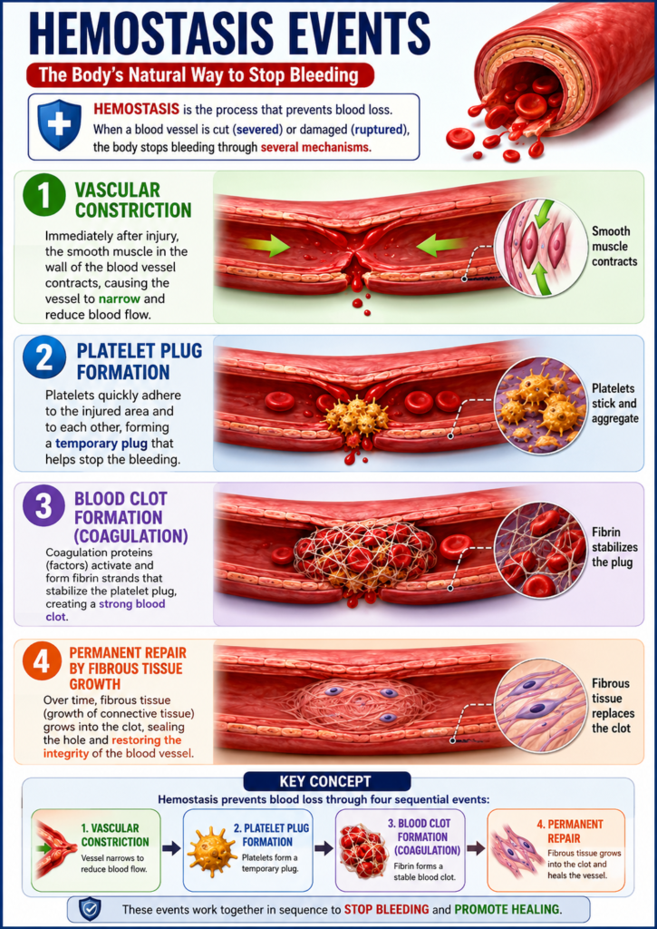
- Immediately after a blood vessel is cut or ruptured, the injury to the vessel wall causes the smooth muscle in the wall to contract.
- This contraction quickly reduces the flow of blood from the damaged blood vessel.
- The contraction occurs because of three factors:
- (1) Local myogenic spasm
- (2) Local autacoid factors released from:
- Traumatized tissues
- Vascular endothelium
- Blood platelets
- (3) Nervous reflexes
- Nervous reflexes start because of:
- Pain nerve impulses, or
- Other sensory impulses coming from the injured blood vessel or nearby tissues.
- However, most vasoconstriction probably occurs because of local myogenic contraction.
- This contraction is triggered directly by damage to the blood vessel wall.
- In smaller blood vessels, platelets are mainly responsible for vasoconstriction.
- Platelets cause this effect by releasing a vasoconstrictor substance called thromboxane A₂.
- The more severe the injury to the blood vessel, the greater the vascular spasm.
- The vascular spasm can continue for many minutes or even hours.
- During this time:
- Platelet plug formation takes place.
- Blood coagulation (clot formation) takes place.
Key Concept
- Vascular constriction is the first step of hemostasis.
- It reduces blood loss immediately by narrowing the damaged blood vessel through local myogenic spasm, autacoid factors, nervous reflexes, and platelet-released thromboxane A₂.
FORMATION OF THE PLATELET PLUG
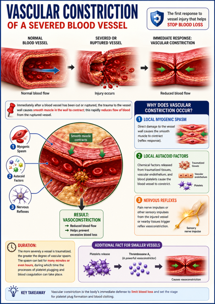
- If the cut in a blood vessel is very small, it is often closed by a platelet plug instead of a blood clot.
- Many tiny holes develop in blood vessels throughout the body every day.
- These small vascular holes are normally sealed by platelets.
Key Concept
- Platelets seal small blood vessel injuries by forming a platelet plug, often without the need for a blood clot.
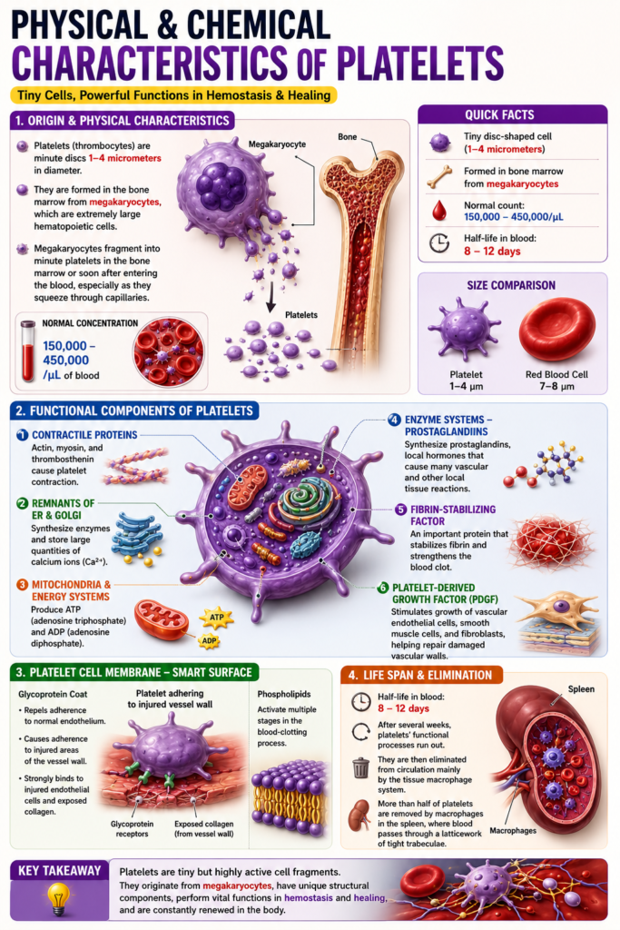
Physical and Chemical Characteristics of Platelets
- Platelets (also called thrombocytes) are very small disc-shaped cells.
- They are 1 to 4 micrometers in diameter.
- Platelets are formed in the bone marrow from megakaryocytes.
- Megakaryocytes are very large blood-forming (hematopoietic) cells in the bone marrow.
- Megakaryocytes break into many tiny platelets in the bone marrow or soon after entering the bloodstream.
- This fragmentation happens especially when they pass through capillaries.
- The normal platelet count in blood is 150,000 to 450,000/μL.
Functional Characteristics of Platelets
- Platelets have many functions similar to whole cells.
- However, they do not have a nucleus.
- Therefore, they cannot reproduce.
- The platelet cytoplasm contains:
- (1) Actin and myosin proteins
- These are contractile proteins similar to those in muscle cells.
- Platelets also contain thrombosthenin, another contractile protein.
- Thrombosthenin helps platelets contract.
- (2) Remnants of the endoplasmic reticulum and Golgi apparatus
- They produce different enzymes.
- They store large amounts of calcium ions.
- (3) Mitochondria and enzyme systems
- They produce adenosine triphosphate (ATP).
- They also produce adenosine diphosphate (ADP).
- (4) Enzyme systems that produce prostaglandins
- Prostaglandins are local hormones.
- They cause many reactions in blood vessels and nearby tissues.
- (5) Fibrin-stabilizing factor
- It is an important protein involved in blood coagulation.
- (6) Platelet-derived growth factor
- It stimulates vascular endothelial cells to grow.
- It stimulates vascular smooth muscle cells to grow.
- It stimulates fibroblasts to grow.
- This cell growth helps repair damaged blood vessel walls.
- (1) Actin and myosin proteins
Platelet Cell Membrane
- The platelet surface is covered by a glycoprotein coat.
- This glycoprotein coat:
- Prevents platelets from sticking to normal endothelium.
- Helps platelets stick to injured areas of the blood vessel wall.
- It especially attaches to injured endothelial cells.
- It attaches even more strongly to exposed collagen in the deeper vessel wall.
- The platelet membrane also contains phospholipids.
- These phospholipids activate several steps of the blood-clotting process.
Platelet Life Span
- Platelets are active structures.
- Their half-life in the blood is 8 to 12 days.
- After several weeks, their functions gradually stop.
- They are then removed from the bloodstream mainly by the tissue macrophage system.
- More than half of the platelets are removed by macrophages in the spleen.
- This occurs as blood passes through the tight trabecular network of the spleen.
Key Concept
- Platelets are small, nucleus-free cell fragments formed from megakaryocytes. They contain contractile proteins, enzymes, ATP, ADP, prostaglandins, fibrin-stabilizing factor, and platelet-derived growth factor. Their membrane helps them attach to injured vessels and supports blood clotting, while macrophages—mainly in the spleen—remove old platelets after a half-life of 8–12 days.
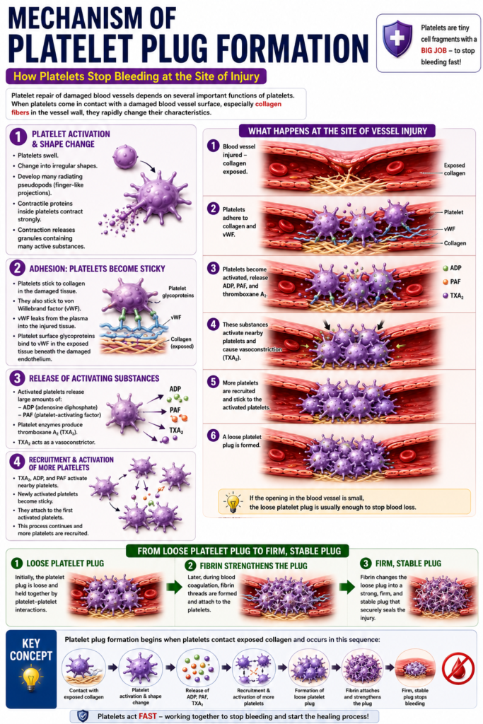
Mechanism of Platelet Plug Formation
- Platelet repair of damaged blood vessels depends on several important functions of platelets.
- When platelets come in contact with a damaged blood vessel surface, especially collagen fibers in the vessel wall, they rapidly change their characteristics.
- Figure 37.1 shows these changes.
- Platelets begin to swell.
- They change into irregular shapes.
- They develop many radiating pseudopods (finger-like projections) on their surface.
- The contractile proteins inside platelets contract strongly.
- This contraction releases granules containing many active substances.
- Platelets become sticky.
- They stick to collagen in the damaged tissue.
- They also stick to a protein called von Willebrand factor (vWF).
- vWF leaks from the plasma into the injured tissue.
- Platelet surface glycoproteins bind to vWF in the exposed tissue beneath the damaged endothelium.
- Activated platelets release larger amounts of adenosine diphosphate (ADP).
- They also release platelet-activating factor (PAF).
- Platelet enzymes produce thromboxane A₂.
- Thromboxane A₂ acts as a vasoconstrictor.
- Thromboxane A₂, ADP, and PAF activate nearby platelets.
- These newly activated platelets become sticky.
- They attach to the first activated platelets.
- At the site of the blood vessel injury, the damaged vessel wall activates more and more platelets.
- These activated platelets attract additional platelets.
- This process forms a platelet plug.
- At first, the platelet plug is loose.
- If the opening in the blood vessel is small, the loose platelet plug is usually enough to stop blood loss.
- Later, during blood coagulation, fibrin threads are formed.
- Fibrin threads attach tightly to the platelets.
- This changes the loose platelet plug into a strong, firm, and stable plug.
Key Concept
- Platelet plug formation begins when platelets contact exposed collagen. Platelets become activated, change shape, become sticky, release ADP, PAF, and thromboxane A₂, recruit more platelets, and form a loose platelet plug. Later, fibrin strengthens the plug into a firm and stable seal.
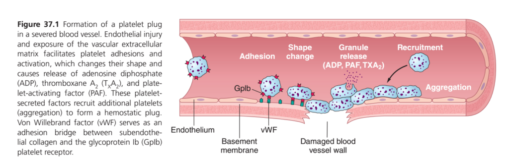
Formation of a Platelet Plug (Guyton Figure 37.1) — SUPERFAST Summary
One-Line Concept
When a blood vessel is injured, platelets act like emergency repair workers. They quickly stick to the damaged area, become activated, call more platelets, and form a temporary plug to stop bleeding.
Step 1: Blood Vessel Injury
🩸 What happens?
- The inner lining (endothelium) is damaged.
- The underlying collagen and basement membrane become exposed.
Concept
➡️ Damage exposes the “sticky surface” that platelets recognize.
Step 2: Platelet Adhesion (First Platelet Sticks)
🟦 Platelets arrive at the injury.
They cannot stick directly very well, so they use a helper protein:
von Willebrand Factor (vWF)
vWF acts like double-sided glue.
- One side binds exposed collagen.
- The other side binds the platelet receptor GPIb (GpIb).
Easy Memory
vWF = Velcro Glue
- Collagen ← vWF → Platelet (GpIb)
Result
✅ First platelet becomes attached.Step 3: Platelet Shape Change
Once attached, the platelet becomes activated.
It changes from:
🔵 Smooth, round platelet
➡️
⭐ Spiky platelet with long projections (pseudopods)
Why?
The spikes help it:
- Stick more firmly
- Cover more damaged surface
- Catch other platelets
Step 4: Granule Release (Chemical Alarm)
Activated platelets release chemicals from their granules.
Important chemicals:
- ADP
- TXA₂ (Thromboxane A₂)
- PAF (Platelet Activating Factor)
Easy Concept
Think of these chemicals as an emergency WhatsApp message saying:
“Everyone come here! Blood vessel is damaged!”
Step 5: Recruitment
The released chemicals attract many more platelets.
More and more platelets arrive at the injury.
This process is called:
Platelet Recruitment
Step 6: Platelet Aggregation
The newly arrived platelets stick to each other.
This forms a large platelet plug.
Purpose
✔ Covers the damaged blood vessel.
✔ Stops blood from leaking.
This plug is called the primary hemostatic plug.
Flow Chart
Blood vessel injury
        ↓
Collagen exposed
        ↓
vWF attaches collagen
        ↓
Platelet binds through GPIb
        ↓
Platelet activation
        ↓
Shape change (spiky platelet)
        ↓
Release ADP + TXA₂ + PAF
        ↓
Recruit more platelets
        ↓
Platelet aggregation
        ↓
Temporary platelet plug formed
Easy Story (Remember Forever)
Imagine there is a hole in a road.
🚧 1. Road breaks → Blood vessel injury
👷 2. First worker arrives → Platelet adhesion
🪢 3. Worker uses rope (vWF) → Platelet sticks to collagen
⭐ 4. Worker opens his arms (shape change) → Better grip
📢 5. Worker shouts for help (ADP, TXA₂, PAF)
👷👷👷 6. More workers arrive (recruitment)
🤝 7. Workers hold hands (aggregation)
🛠 8. Hole is temporarily covered (platelet plug)
High-Yield Exam Points
| Step | Key Event | Important Molecule |
|---|---|---|
| 1 | Vessel injury | Collagen exposed |
| 2 | Adhesion | vWF + GPIb |
| 3 | Activation | Shape change, pseudopods |
| 4 | Granule release | ADP, TXA₂, PAF |
| 5 | Recruitment | More platelets attracted |
| 6 | Aggregation | Platelet plug formation |
Mnemonic
“A SMART Plug”
- A = Adhesion
- S = Shape change
- M = Mediator release (ADP, TXA₂, PAF)
- R = Recruitment
- A = Aggregation
- T Plug = Temporary Platelet Plug
Final Concept in One Sentence
After blood vessel injury, platelets stick to exposed collagen through vWF–GPIb, become activated and change shape, release ADP, TXA₂, and PAF to recruit more platelets, and aggregate together to form a temporary platelet plug that stops bleeding.
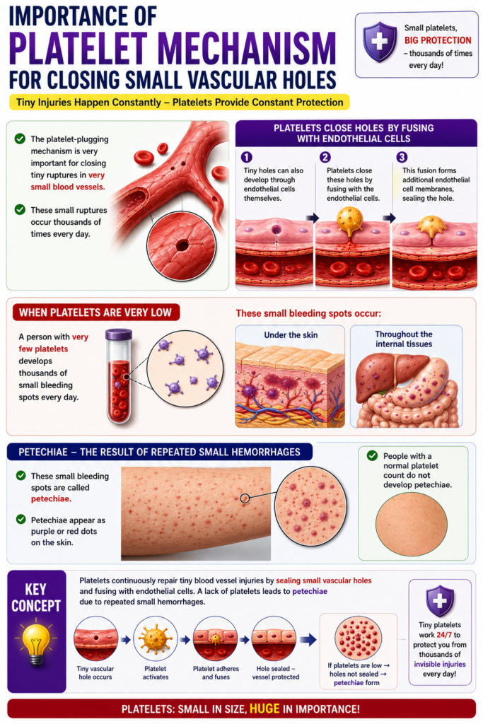
Importance of Platelet Mechanism for Closing Small Vascular Holes
- The platelet-plugging mechanism is very important for closing tiny ruptures in very small blood vessels.
- These small ruptures occur thousands of times every day.
- Many tiny holes can also develop through the endothelial cells themselves.
- Platelets close these holes by fusing with the endothelial cells.
- This fusion forms additional endothelial cell membranes.
- A person with very few platelets develops thousands of small bleeding spots every day.
- These small bleeding spots occur:
- Under the skin
- Throughout the internal tissues
- These spots are called petechiae.
- Petechiae appear as purple or red dots on the skin.
- People with a normal platelet count do not develop petechiae.
Key Concept
- Platelets continuously repair tiny blood vessel injuries by sealing small vascular holes and fusing with endothelial cells. A lack of platelets leads to petechiae due to repeated small hemorrhages.
BLOOD COAGULATION IN THE RUPTURED VESSEL
- The third mechanism of hemostasis is blood clot formation.
- If the injury to the blood vessel wall is severe, the blood clot begins to form within 15–20 seconds.
- If the injury is minor, the blood clot begins to form within 1–2 minutes.
- Clotting starts because of activator substances released from:
- The injured blood vessel wall
- Platelets
- Blood proteins attached to the injured vessel wall
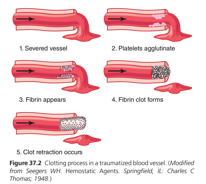
- Figure 37.2 shows the physical events of the blood clotting process.
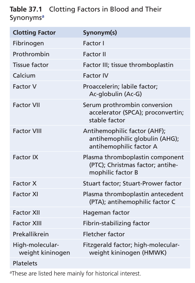
- Table 37.1 lists the most important clotting factors.
- Within 3–6 minutes after a blood vessel ruptures, the entire opening or broken end is filled with a blood clot, if the opening is not too large.
- After 20–60 minutes, the blood clot retracts (shrinks).
- Clot retraction further closes the blood vessel.
- Platelets play an important role in clot retraction.
Key Concept
- Blood coagulation is the third step of hemostasis. Activator substances from the injured vessel wall, platelets, and blood proteins start clot formation. The clot fills the vessel opening within 3–6 minutes and later retracts to further reduce bleeding.
Clotting Process in a Traumatized Blood Vessel (Guyton Figure 37.2) — Superfast conceptual Summary
One-Line Concept
After a blood vessel is injured, the body first makes a temporary platelet plug, then strengthens it with fibrin threads, and finally tightens the clot to permanently stop bleeding.
Step 1: Severed Blood Vessel 🩸
What happens?
- The blood vessel is cut or damaged.
- Blood starts leaking out.
Concept
Think of it as a burst water pipe that needs immediate repair.
Goal: Stop blood loss as quickly as possible.Step 2: Platelets Agglutinate (Primary Plug) 🟣
What happens?
Platelets rush to the injured site and stick together.
This forms a temporary platelet plug.
Easy Concept
Imagine workers arriving quickly and standing shoulder-to-shoulder to block the hole.
Important Point
This plug is:
- Fast
- Temporary
- Not very strong
Purpose
✔ Slows bleeding immediately.
Step 3: Fibrin Appears 🕸️
What happens?
The coagulation (clotting) cascade becomes activated.
It converts:
Fibrinogen (soluble protein)
⬇️
Fibrin (insoluble protein threads)
These fibrin threads begin to appear around the platelet plug.
Easy Concept
Think of fibrin as a strong fishing net thrown over the platelet plug.
Step 4: Fibrin Clot Forms 🧱
What happens?
More fibrin strands are produced.
They form a mesh that traps:
- Platelets
- Red blood cells (RBCs)
- White blood cells (WBCs)
Everything becomes locked together.
Easy Concept
Imagine pouring cement over bricks.
The platelets are the bricks.
The fibrin is the cement.
Now the repair becomes much stronger.
Result
✔ Strong blood clot formed.
✔ Bleeding stops completely.
Step 5: Clot Retraction 🧲
What happens?
Platelets contract using contractile proteins inside them.
They pull on the fibrin threads.
The clot becomes:
- Smaller
- Tighter
- Stronger
The edges of the damaged blood vessel move closer together.
Easy Concept
Like pulling the drawstrings of a bag.
The opening becomes smaller and closes.
Benefits
✔ Stronger clot
✔ Less bleeding
✔ Faster healing
Complete Flow
Blood vessel injury
        ↓
Platelets gather
        ↓
Temporary platelet plug
        ↓
Fibrin appears
        ↓
Fibrin mesh forms
        ↓
Strong fibrin clot
        ↓
Clot retracts (tightens)
        ↓
Bleeding stops and healing begins
Easy Story (Remember Forever)
Imagine a wall with a hole.
🧱 1. Wall breaks → Blood vessel injury
👷 2. Workers arrive → Platelets gather
🪵 3. Wooden boards cover the hole → Temporary platelet plug
🕸️ 4. Steel wires are added → Fibrin appears
🏗️ 5. Concrete hardens everything → Fibrin clot forms
🤲 6. Workers tighten the repair → Clot retraction
Finally, the wall becomes strong and ready for permanent repair.
High-Yield Exam Table
| Step | Event | Main Function |
|---|---|---|
| 1 | Vessel injury | Blood vessel is damaged |
| 2 | Platelet agglutination | Temporary platelet plug forms |
| 3 | Fibrin appears | Clotting cascade produces fibrin threads |
| 4 | Fibrin clot forms | Stable clot traps blood cells and platelets |
| 5 | Clot retraction | Clot shrinks, strengthens, and pulls wound edges together |
Easy Mnemonic
“PFCR”
- P = Platelets plug
- F = Fibrin appears
- C = Clot forms
- R = Retraction
Difference Between Platelet Plug and Fibrin Clot
| Platelet Plug | Fibrin Clot |
|---|---|
| Forms first | Forms after platelet plug |
| Temporary | Permanent and stable |
| Made mainly of platelets | Made of fibrin + platelets + trapped blood cells |
| Stops bleeding quickly | Completely seals the damaged vessel |
Final Concept in One Sentence
When a blood vessel is injured, platelets rapidly form a temporary plug to reduce bleeding, fibrin threads then reinforce the plug into a strong and stable clot, and finally the clot retracts, pulling the wound edges together to stop bleeding and promote healing.
FIBROUS ORGANIZATION OR DISSOLUTION OF BLOOD CLOTS
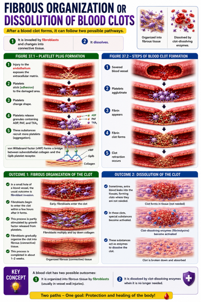
- After a blood clot forms, it can follow two possible pathways:
- (1) It is invaded by fibroblasts and changes into connective tissue.
- (2) It dissolves.
- Figure 37.1 shows platelet plug formation:
- Injury to the endothelium exposes the extracellular matrix.
- Platelets stick (adhesion) to the damaged area.
- Platelets change shape.
- Platelets release granules containing ADP, PAF, and TXA₂.
- These substances recruit more platelets (aggregation).
- von Willebrand factor (vWF) forms a bridge between subendothelial collagen and the GpIb platelet receptor.
- Figure 37.2 shows the steps of blood clot formation:
- 1. Severed blood vessel
- 2. Platelets agglutinate
- 3. Fibrin appears
- 4. Fibrin clot forms
- 5. Clot retraction occurs
- In a small hole of a blood vessel, the usual outcome is fibroblast invasion.
- Fibroblasts begin to enter the clot within a few hours after it forms.
- This process is partly stimulated by growth factor released from platelets.
- Fibroblasts gradually organize the clot into fibrous (connective) tissue.
- This process is completed in about 1–2 weeks.
- Sometimes, extra blood leaks into the tissues.
- This forms tissue clots where they are not needed.
- In these clots, special substances become activated.
- These substances act as enzymes to dissolve the clot.
Key Concept
- A blood clot has two possible outcomes: it is either organized into fibrous tissue by fibroblasts (usually in vessel wall injuries) or dissolved by clot-dissolving enzymes when it is no longer needed.
MADE BY SELF LEARNING CEO AND FOUNDER DR SHEEN
