


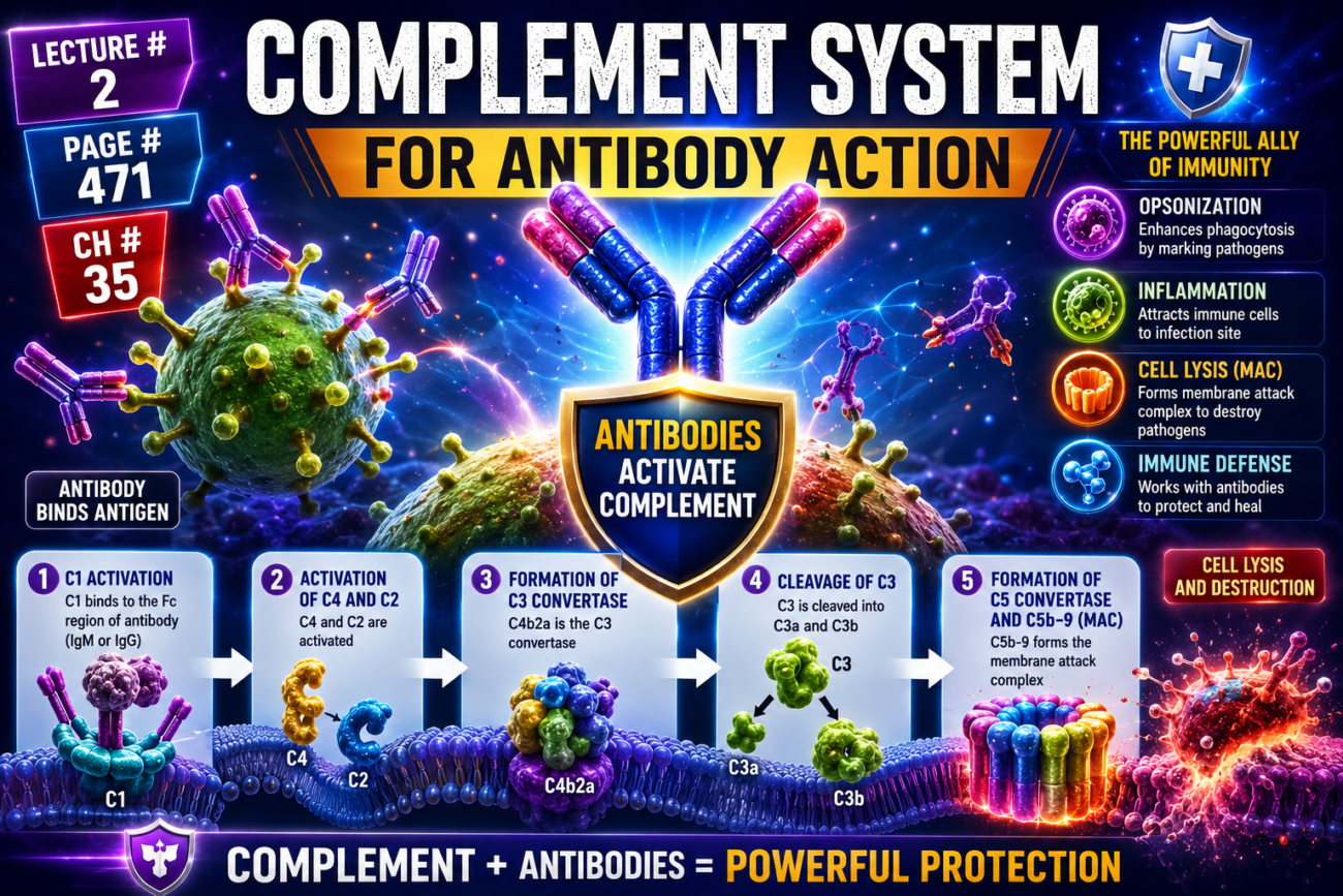
- The main function of the complement system is to enhance (complement) the actions of:
- Antibodies
- Phagocytic cells
- The complement system helps:
- Neutralize pathogens.
- Destroy pathogens.
- Remove damaged cells from the body.
- Promote inflammation.
- Complement is the collective name for a system of about 20 proteins.
- Many of these proteins are enzyme precursors.
- The principal proteins of the complement system are:
- C1
- C2
- C3
- C4
- C5
- C6
- C7
- C8
- C9
- Factor B
- Factor D
- These complement proteins are normally present among the plasma proteins in the blood.
- They are also present among the proteins that leak from capillaries into the tissue spaces.
- The complement proteins normally remain inactive.
- They become activated through the classical pathway.
Figure Number
- Figure 35.6: Shows the complement system proteins (C1–C9, Factor B, and Factor D) and their activation through the classical pathway.
Key Concept
The complement system is a group of about 20 plasma proteins that enhance the actions of antibodies and phagocytic cells. It helps destroy pathogens, remove damaged cells, and promote inflammation. The major complement proteins (C1–C9, Factor B, and Factor D) circulate in an inactive form and are activated through the classical pathway.
Classical Pathway of the Complement System
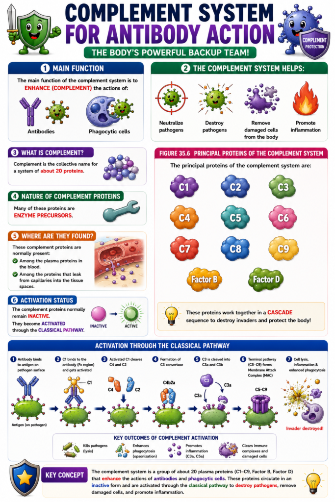
- The classical pathway begins with an antigen-antibody reaction.
- When an antibody binds to an antigen, a reactive site on the constant region of the antibody becomes exposed (activated).
- This activated site binds directly to the C1 protein of the complement system.
- Binding to C1 starts a cascade of sequential reactions.
- The process begins with activation of the C1 proenzyme.
- Activated C1 enzymes successively activate increasing amounts of complement enzymes.
- As the cascade continues, the response becomes greatly amplified.
- Multiple complement end products are formed.
- These end products help protect the body’s tissues from damage caused by invading organisms or toxins.
- The major effects of complement activation are:
- 1. Opsonization and Phagocytosis
- C3b strongly enhances phagocytosis by neutrophils and macrophages.
- These phagocytes engulf bacteria coated with antigen-antibody complexes.
- This process is called opsonization.
- Opsonization can increase bacterial destruction many hundredfold.
- 2. Lysis
- The membrane attack complex (MAC) is one of the most important complement products.
- The MAC consists of C5b, C6, C7, C8, and C9 (C5b6789).
- It inserts into the lipid bilayer of the cell membrane.
- It forms pores that allow ions to pass.
- Water enters the cell by osmosis.
- This causes osmotic rupture (lysis) of bacteria and other invading organisms.
- 3. Agglutination
- Complement products alter the surface of invading organisms.
- This causes microorganisms to stick together.
- The result is agglutination.
- 4. Neutralization of Viruses
- Complement enzymes and other complement products attack the structure of some viruses.
- This makes the viruses nonvirulent.
- 5. Chemotaxis
- C5a attracts neutrophils and macrophages to the infected tissue.
- This movement of phagocytes toward the infection is called chemotaxis.
- 6. Activation of Mast Cells and Basophils
- C3a, C4a, and C5a activate mast cells and basophils.
- These cells release:
- Histamine
- Heparin
- Several other chemical substances
- These substances cause:
- Increased local blood flow
- Increased leakage of plasma proteins and fluid into tissues
- Local tissue reactions that help immobilize or destroy the antigen.
- These reactions also contribute to inflammation and allergy.
- 7. Inflammatory Effects
- Complement products further increase the inflammatory response.
- They cause:
- Further increase in local blood flow.
- Greater leakage of proteins from capillaries.
- Coagulation of interstitial fluid proteins within tissue spaces.
- This helps prevent the invading organism from spreading through the tissues.
Figure Number
- Figure 35.6: Shows:
- Activation of the classical complement pathway
- Sequential activation of complement proteins beginning with C1
- Formation of complement end products, including C3b and the membrane attack complex (C5b6789)
- Major biological effects of complement activation
Key Concept
The classical complement pathway is triggered when an antigen-antibody complex activates C1, initiating an amplified enzyme cascade. The resulting complement products produce opsonization (C3b), membrane attack complex formation (C5b–C9), agglutination, viral neutralization, chemotaxis (C5a), mast cell and basophil activation (C3a, C4a, C5a), and powerful inflammatory responses that together eliminate pathogens and limit their spread.
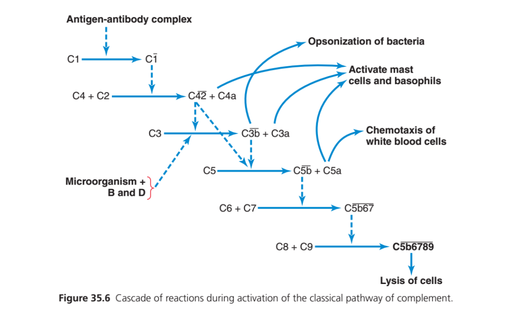
Guyton Physiology Figure 35.6 Explained in Detail
Complement System – Classical Pathway (Cascade of Complement Activation)
This is one of the most important and highest-yield figures in immunology. It explains how antibodies activate the complement system, resulting in:
- Opsonization (enhanced phagocytosis)
- Inflammation
- Chemotaxis of leukocytes
- Mast cell activation
- Direct killing (lysis) of microorganisms
This figure answers an important question:
“After antibodies bind to bacteria, how are the bacteria actually destroyed?”
The answer is:
Through activation of the Complement System.
Overall Concept
Imagine a thief enters a building.
One security guard (antibody) catches him.
But the security guard alone cannot remove the thief.
Instead,
he presses an emergency alarm.
Immediately,
- Police arrive
- Ambulance arrives
- Fire brigade arrives
- Special forces arrive
One alarm activates many different emergency teams.
The complement system works exactly like this.
The antibody simply starts the alarm.
Complement proteins perform the destruction.
What is the Complement System?
The complement system is a group of approximately 30 plasma proteins (Guyton often emphasizes the major proteins C1–C9).
These proteins normally circulate in the blood as inactive precursors (zymogens).
When activated, they trigger a cascade reaction, where one activated protein activates the next.
Why is it called “Complement”?
Because it complements (enhances) the action of antibodies.
Antibodies recognize the pathogen.
Complement helps eliminate it.
First Look at the Figure
The figure shows the Classical Complement Pathway.
It begins with
Antigen–Antibody Complex
and ends with
Lysis of the Cell
Notice that each step activates the next.
This is called a
Cascade Reaction
What is a Cascade?
Think of a row of dominoes.
Domino 1
   ↓
Domino 2
   ↓
Domino 3
   ↓
Domino 4
Push one domino,
and every other domino falls.
Exactly the same thing happens in the complement system.
One activated complement protein activates the next one.
Step 1
Antigen–Antibody Complex
Look at the top left.
Everything starts here.
What is an Antigen–Antibody Complex?
An antibody (usually IgG or IgM) binds to an antigen on the surface of a microorganism.
This binding exposes the Fc (constant) region of the antibody.
The exposed Fc region allows C1 to bind.
Example
A bacterium enters the body.
↓
IgG antibody binds to the bacterial surface.
↓
This forms an antigen–antibody complex.
↓
Now complement activation begins.
Step 2
Activation of C1
The first complement protein is
C1
Normally,
C1 is inactive.
When it binds to the Fc region of antibody,
it becomes
Activated C1 (C1̅)
This is the first enzyme in the classical pathway.
Why is C1 Important?
C1 starts the entire cascade.
Without C1,
the classical pathway cannot proceed.
Easy Analogy
Imagine turning on the main switch in a factory.
Until the switch is turned on,
no machine works.
C1 is that master switch.
Step 3
Activation of C4 and C2
Activated C1 cleaves:
- C4 → C4a + C4b
- C2 → C2a + C2b
In Guyton’s notation, the active enzyme complex is shown as C42, which functions as the C3 convertase of the classical pathway.
Function of C3 Convertase
Its only job is to activate
C3
Step 4
Activation of C3
This is the most important step in the complement cascade.
C3 is the most abundant complement protein.
C3 convertase splits C3 into:
- C3a
- C3b
Each fragment has different functions.
C3b
Opsonization
One arrow points toward
Opsonization of Bacteriahat is Opsonization?
Opsonization means:
Coating bacteria to make them easier for phagocytes to recognize and ingest.
C3b binds to the microbial surface.
Neutrophils and macrophages have C3b receptors.
They attach more easily and phagocytose the bacterium.
Easy Analogy
Imagine a criminal wearing a bright fluorescent jacket.
Police can spot and catch him much more easily.
C3b acts like that fluorescent marker.
Clinical Example
People with C3 deficiency experience recurrent bacterial infections because bacteria are not efficiently opsonized and phagocytosed.
C3a
Activation of Mast Cells and Basophils
Another arrow points toward
Activate Mast Cells and Basophils
What Happens?
C3a stimulates mast cells and basophils to release:
- Histamine
- Other inflammatory mediators
Effects of Histamine
- Vasodilation
- Increased vascular permeability
- Recruitment of immune cells
This enhances inflammation.
Easy Analogy
Think of C3a as an emergency siren that tells nearby inflammatory cells to release chemical signals.
Step 5
Activation of C5
The figure shows that C5 is cleaved into:
- C5a
- C5b
These fragments have different functions.
C5a
Chemotaxis
One arrow points toward
Chemotaxis of White Blood Cells
What is Chemotaxis?
Chemotaxis is the directed movement of leukocytes toward higher concentrations of chemical signals.
C5a is one of the most powerful chemotactic factors in the complement system.
Which Cells Respond?
- Neutrophils
- Monocytes
- Macrophages
These cells migrate toward the site of infection.
Easy Analogy
Imagine the smell of food becoming stronger as you approach the kitchen.
Leukocytes “follow the smell” of C5a toward the infection.
C5a Also Activates Mast Cells
C5a is an even more potent anaphylatoxin than C3a.
It stimulates mast cells and basophils to release histamine, further amplifying inflammation.
Step 6
Formation of C5b67
C5b binds sequentially with:
- C6
- C7
forming
C5b67
This complex attaches firmly to the microbial membrane.
Function
It prepares the membrane for the insertion of additional complement proteins.
Analogy
Imagine workers constructing the frame of a tunnel before opening the passage.
Step 7
Addition of C8 and C9
Next,
C8 and multiple C9 molecules bind.
This forms:
C5b6789
This structure is known as the
Membrane Attack Complex (MAC)
Membrane Attack Complex (MAC)
The MAC creates a pore (hole) in the microbial cell membrane.
Water and ions rush through the pore.
The cell loses membrane integrity and bursts.
This process is called:
Cell Lysis
Easy Analogy
Imagine puncturing a water balloon.
Water escapes,
the balloon collapses.
Similarly,
the microorganism is destroyed.
Alternative Pathway (Shown on the Left)
Notice another arrow labeled:
Microorganism + B and D
This represents the Alternative Complement Pathway.
Unlike the classical pathway,
it does not require antibodies.
Instead,
microbial surfaces directly activate complement with the help of Factors B and D.
Both the classical and alternative pathways converge at C3 activation, after which they share the same downstream events leading to opsonization, inflammation, and MAC formation.
Functions of Major Complement Components
| Complement Component | Main Function |
|---|---|
| C1 | Initiates the classical pathway after binding antigen–antibody complexes |
| C3b | Opsonization of microorganisms |
| C3a | Mast cell activation and inflammation (anaphylatoxin) |
| C5a | Powerful chemotaxis and mast cell activation |
| C5b | Initiates membrane attack complex formation |
| C6, C7, C8, C9 | Form the membrane attack complex (MAC) |
| MAC (C5b-9) | Direct lysis of susceptible cells |
Clinical Correlations
1. Neisseria Infections
Deficiency of terminal complement components (C5–C9) impairs MAC formation.
Patients are particularly susceptible to recurrent infections with Neisseria meningitidis and Neisseria gonorrhoeae.
2. C3 Deficiency
C3 deficiency causes severe recurrent bacterial infections because both opsonization and downstream complement activation are impaired.3. Hereditary Angioedema
Deficiency of C1 esterase inhibitor leads to uncontrolled activation of the classical pathway and excessive bradykinin production, causing recurrent episodes of severe swelling.
4. Paroxysmal Nocturnal Hemoglobinuria (PNH)
Red blood cells lack protective complement-regulating proteins (CD55 and CD59).
Complement forms the MAC on RBCs, causing intravascular hemolysis.
Complete Flow Chart
Antigen + IgG/IgM Antibody
            ↓
Antigen–Antibody Complex
            ↓
C1 Activation
            ↓
C4 + C2 Activation
            ↓
C3 Convertase Formation
            ↓
C3 → C3a + C3b
        ↓         ↓
 Mast Cell     Opsonization
 Activation
            ↓
C5 → C5a + C5b
      ↓         ↓
 Chemotaxis   C6 + C7
              ↓
            C5b67
              ↓
           C8 + C9
              ↓
Membrane Attack Complex (MAC)
              ↓
Pore Formation
              ↓
Lysis of the Microorganism
Connecting Figures 35.1–35.6
| Figure | Main Concept |
|---|---|
| Figure 35.1 | Development of T and B lymphocytes. |
| Figure 35.2 | Clonal selection and activation of B cells. |
| Figure 35.3 | Primary and secondary antibody responses. |
| Figure 35.4 | Structure of the IgG antibody. |
| Figure 35.5 | Antibody-mediated agglutination of antigens. |
| Figure 35.6 | Activation of the complement system by antigen–antibody complexes, leading to inflammation, opsonization, chemotaxis, and direct microbial lysis. |
Together, these figures describe the progression of humoral immunity:
B-cell development → Antibody production → Antigen binding → Agglutination → Complement activation → Pathogen destruction.
High-Yield MBBS Pearls
- The classical pathway is activated by IgG or IgM bound to antigen.
- C1 initiates the classical complement cascade.
- C3 is the central complement protein and a major point of amplification.
- C3b = Opsonization (“b = bacteria coated”).
- C3a and C5a = Anaphylatoxins, promoting mast cell degranulation and inflammation.
- C5a = Powerful chemotactic factor for neutrophils and monocytes.
- C5b–C9 = Membrane Attack Complex (MAC), which forms pores and lyses susceptible cells.
- The alternative pathway does not require antibodies and is activated directly by microbial surfaces.
Key Concept
Figure 35.6 illustrates the classical complement pathway, a powerful amplification system that is triggered when antibodies bind to an antigen. Activation of C1 initiates a cascade culminating in C3 activation, which produces C3b for opsonization and C3a for inflammation. C5a recruits leukocytes by chemotaxis and further enhances inflammation, while C5b initiates assembly of the membrane attack complex (C5b–9), creating pores in microbial membranes and causing direct cell lysis. Thus, complement transforms antibody binding into efficient pathogen elimination through multiple coordinated mechanisms.
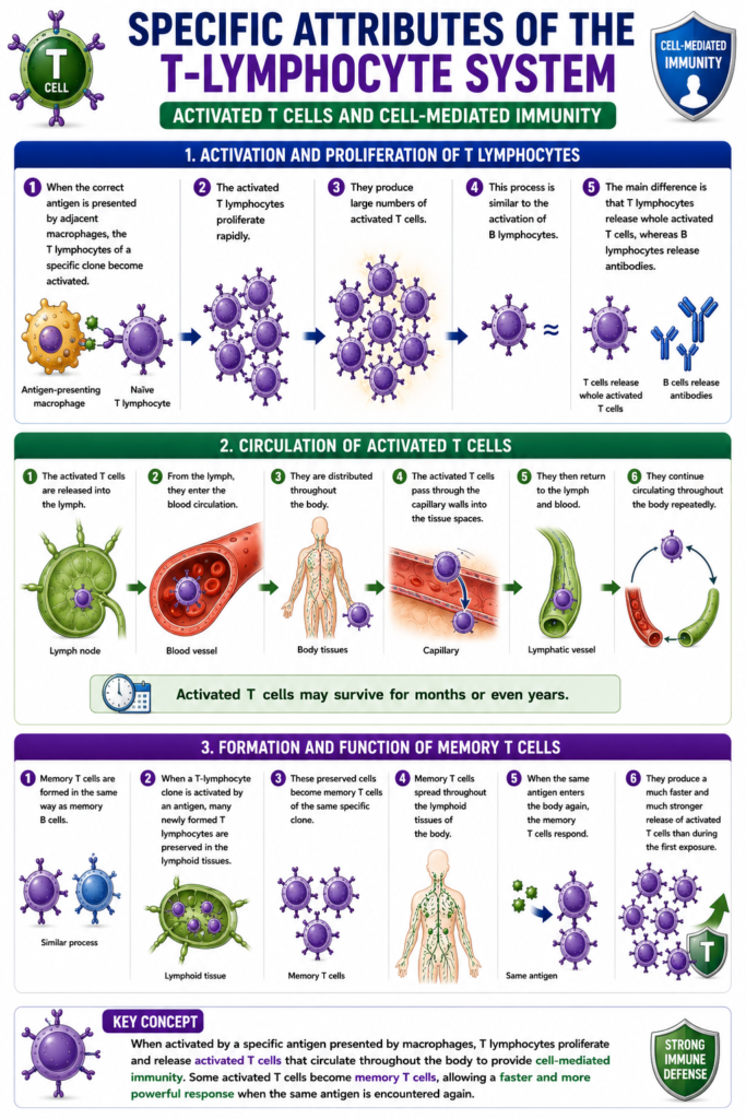
Specific Attributes of the T-Lymphocyte System—Activated T Cells and Cell-Mediated Immunity
- When the correct antigen is presented by adjacent macrophages, the T lymphocytes of a specific clone become activated.
- The activated T lymphocytes proliferate rapidly.
- They produce large numbers of activated T cells.
- This process is similar to the activation of B lymphocytes.
- The main difference is that T lymphocytes release whole activated T cells, whereas B lymphocytes release antibodies.
- The activated T cells are released into the lymph.
- From the lymph, they enter the blood circulation.
- They are distributed throughout the body.
- The activated T cells pass through the capillary walls into the tissue spaces.
- They then return to the lymph and blood.
- They continue circulating throughout the body repeatedly.
- Activated T cells may survive for months or even years.
- Memory T cells are formed in the same way as memory B cells.
- When a T-lymphocyte clone is activated by an antigen, many newly formed T lymphocytes are preserved in the lymphoid tissues.
- These preserved cells become memory T cells of the same specific clone.
- Memory T cells spread throughout the lymphoid tissues of the body.
- When the same antigen enters the body again, the memory T cells respond.
- They produce a much faster and much stronger release of activated T cells than during the first exposure.
Key Concept
When activated by a specific antigen presented by macrophages, T lymphocytes proliferate and release activated T cells that circulate throughout the body to provide cell-mediated immunity. Some activated T cells become memory T cells, allowing a faster and more powerful response when the same antigen is encountered again.
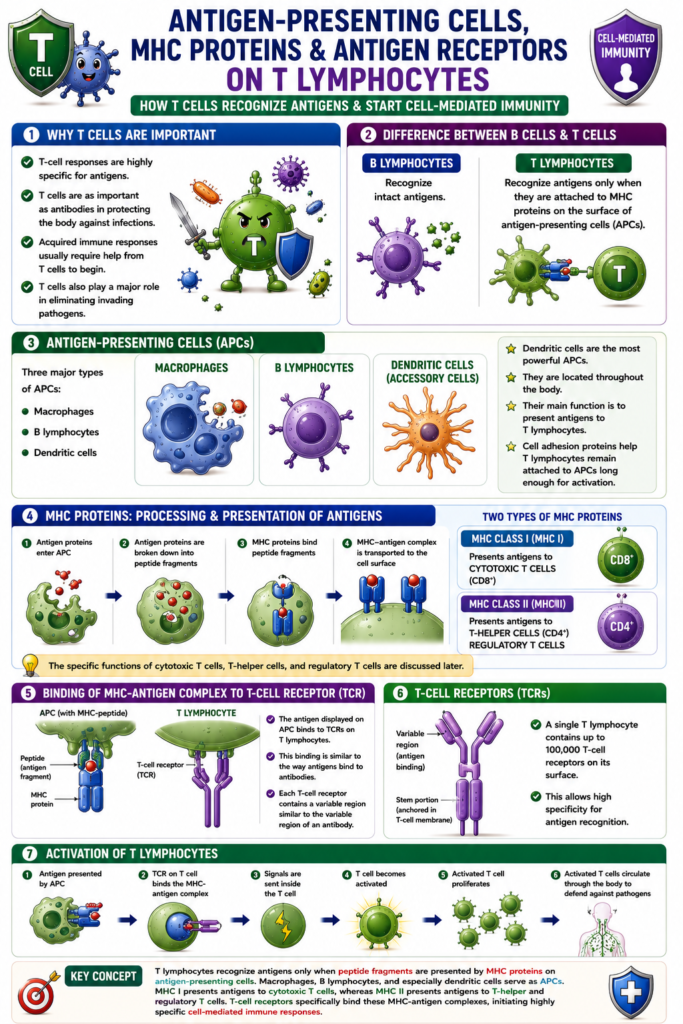
Antigen-Presenting Cells, Major Histocompatibility Complex (MHC) Proteins, and Antigen Receptors on T Lymphocytes
- T-cell responses are highly specific for antigens.
- T-cell responses are as important as antibodies in protecting the body against infections.
- Acquired immune responses usually require help from T cells to begin.
- T cells also play a major role in eliminating invading pathogens.
- B lymphocytes recognize intact antigens.
- T lymphocytes recognize antigens only when they are attached to Major Histocompatibility Complex (MHC) proteins.
- These MHC proteins are present on the surface of antigen-presenting cells (APCs).
- The three major types of antigen-presenting cells are:
- Macrophages
- B lymphocytes
- Dendritic cells
- Dendritic cells are the most powerful antigen-presenting cells.
- Dendritic cells are also called accessory cells.
- They are located throughout the body.
- Their main function is to present antigens to T lymphocytes.
- Cell adhesion proteins help T lymphocytes remain attached to antigen-presenting cells long enough for activation.
- MHC proteins are produced from genes called the Major Histocompatibility Complex (MHC).
- Inside antigen-presenting cells, antigen proteins are broken down into peptide fragments.
- MHC proteins bind these peptide fragments.
- The MHC-antigen complex is transported to the cell surface.
- There are two types of MHC proteins.
- MHC Class I (MHC I):
- Presents antigens to cytotoxic T cells.
- MHC Class II (MHC II):
- Presents antigens to:
- T-helper cells
- Regulatory T cells
- Presents antigens to:
- The specific functions of cytotoxic T cells, T-helper cells, and regulatory T cells are discussed later.
- The antigen displayed on the antigen-presenting cell binds to T-cell receptors (TCRs) on the T lymphocyte.
- This binding is similar to the way antigens bind to antibodies.
- Each T-cell receptor contains a variable region similar to the variable region of an antibody.
- The stem portion of the T-cell receptor is firmly attached to the T-cell membrane.
- A single T lymphocyte contains up to 100,000 T-cell receptors on its surface.
Figure Number
- Figure 35.7: Shows:
- Antigen-presenting cells (macrophages, B lymphocytes, dendritic cells)
- Presentation of antigen peptides by MHC proteins
- Binding of the MHC-antigen complex to T-cell receptors
- Activation of T lymphocytes
Key Concept
T lymphocytes recognize antigens only when peptide fragments are presented by MHC proteins on antigen-presenting cells. Macrophages, B lymphocytes, and especially dendritic cells serve as antigen-presenting cells. MHC I presents antigens to cytotoxic T cells, whereas MHC II presents antigens to T-helper and regulatory T cells. T-cell receptors specifically bind these MHC-antigen complexes, initiating highly specific cell-mediated immune responses.
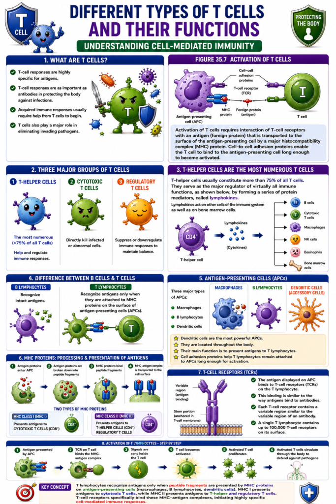
Different Types of T Cells and Their Functions
- There are three major types of T cells.
- These are:
- T-helper cells
- Cytotoxic T cells
- Regulatory T cells (Suppressor T cells)
- Each type of T cell has a different function.
- T-helper cells are the most numerous T cells.
- They make up more than 75% of all T cells.
- As their name suggests, T-helper cells assist other cells of the immune system.
- They act as the major regulators of almost all immune functions.
- T-helper cells produce protein mediators called lymphokines.
- Lymphokines act on:
- Other immune system cells.
- Bone marrow cells.
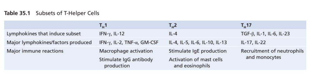
- When naïve CD4⁺ T-helper cells are stimulated, they differentiate into different subsets.
- Each subset produces different lymphokines.
- Each subset performs different immune functions.
- Table 35.1 summarizes:
- The main T-helper cell subsets.
- The lymphokines that induce each subset.
- The lymphokines produced by each subset.
- The immune reactions triggered by each subset.
Figure Number
- Figure 35.7: Shows:
- Activation of T cells
- Binding of the T-cell receptor to an antigen presented by an MHC protein
- Role of cell adhesion proteins in T-cell activation
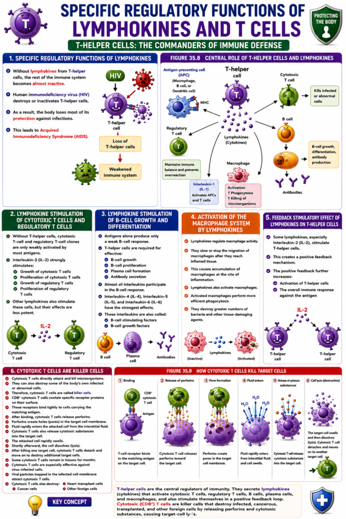
- Figure 35.8: Shows:
- T-helper cells regulating immune functions by producing lymphokines
- Effects of lymphokines on immune cells and bone marrow cells
- Table 35.1: Summarizes:
- Major T-helper cell subsets
- Inducing lymphokines
- Lymphokines produced
- Immune responses triggered by each subset
Key Concept
T cells are classified into T-helper cells, cytotoxic T cells, and regulatory (suppressor) T cells. T-helper cells are the most abundant, accounting for more than 75% of all T cells. They regulate nearly all immune responses by secreting lymphokines that activate immune cells and influence bone marrow function. Activated CD4⁺ T-helper cells differentiate into specialized subsets that produce different lymphokines and initiate specific immune reactions.
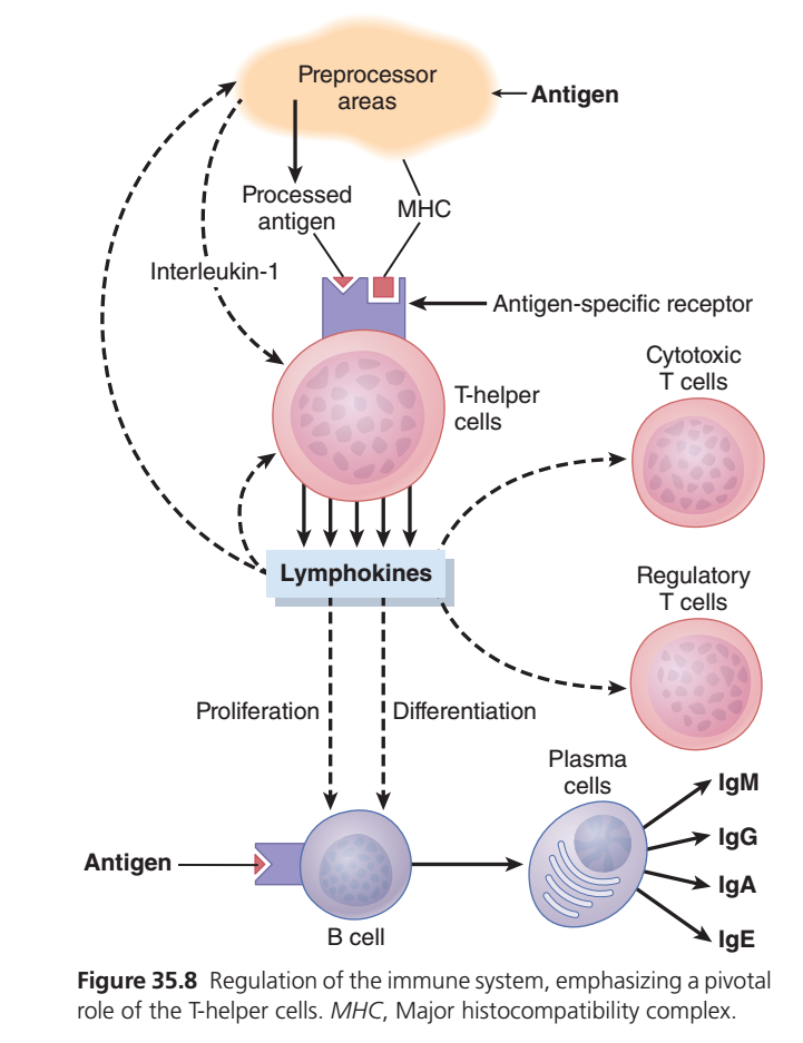
Guyton Physiology Figure 35.8 Explained in Detail
Regulation of the Immune System – Central Role of T-Helper (CD4⁺) Cells
This is one of the most important figures in immunology because it shows that T-helper (CD4⁺) cells are the “master regulators” (or orchestra conductors) of the adaptive immune system.
Without T-helper cells:
- B cells cannot produce antibodies efficiently.
- Cytotoxic T cells are poorly activated.
- Regulatory T cells cannot properly control immunity.
- The immune response becomes weak and uncoordinated.
This is why diseases such as HIV/AIDS, which destroy CD4⁺ T-helper cells, cause severe immunodeficiency.
Overall Concept
Imagine a large army preparing for war.
The army consists of:
- Soldiers → Cytotoxic T cells
- Weapon factories → Plasma cells
- Police officers → Regulatory T cells
- Ammunition factories → B cells
But all of these require orders from the Commander.
The commander is the:
T-Helper Cell (CD4⁺)
Without the commander,
every unit becomes confused.
This figure explains how one activated T-helper cell controls almost the entire immune response.
First Look at the Figure
The figure can be divided into 8 major steps.
Antigen enters body
        ↓
Antigen Presenting Cell (APC)
        ↓
Antigen Processing
        ↓
MHC-II presents antigen
        ↓
T-helper Cell Activation
        ↓
Lymphokine (Cytokine) Release
        ↓
Activation of:
• B cells
• Cytotoxic T cells
• Regulatory T cells
        ↓
Antibody Production + Cell-mediated Immunity
Everything revolves around
The T-helper Cell
PART 1
Antigen
The figure begins with
Antigen
An antigen is
any foreign substance
capable of producing an immune response.
Examples
- Bacteria
- Viruses
- Parasites
- Fungi
- Toxins
Question
Can a T-helper cell recognize a free antigen directly?
No.
Unlike B cells,
T-helper cells cannot recognize free antigen.
The antigen must first be
processed
and
presented.
PART 2
Preprocessor Areas (Antigen-Presenting Cells)
The orange area labeled
Preprocessor Areas
represents
Antigen-Presenting Cells (APCs)
Examples include:
- Dendritic cells (most important)
- Macrophages
- B lymphocytes
What Happens Here?
The APC
engulfs the microorganism.
↓
Breaks it into small peptide fragments.
↓
This is called
Antigen Processing
Easy Analogy
Imagine shredding a large document
into small pages
before showing it to your teacher.
The APC cuts the antigen into pieces before presenting it.
PART 3
Processed Antigen + MHC
After processing,
the peptide fragment is attached to
MHC Class II
(Major Histocompatibility Complex II)
The figure labels
Processed Antigen
MHC
What is MHC?
Think of MHC as
a
Display Tray
or
Presentation Plate.
The APC places
the processed antigen
onto this tray.
Easy Analogy
Imagine a waiter
placing food
onto a serving plate
before taking it to the customer.
The plate is MHC.
The food is antigen.
Which MHC?
For T-helper cells,
the important molecule is
MHC Class II
PART 4
Antigen-Specific Receptor
The T-helper cell has
T-cell Receptor (TCR)
The TCR recognizes
only
one
specific antigen.
Important Concept
The TCR does not recognize free antigen.
It recognizes
Processed Antigen
MHC II
together.
This is called
MHC Restriction
Easy Analogy
Think of a lock that opens only when both the correct key and the correct keychain are present.
The TCR requires:
- the correct peptide (key)
- presented on the correct MHC molecule (keychain)
PART 5
Interleukin-1 (IL-1)
The figure shows
Interleukin-1 (IL-1)
being released by the APC.
Function
IL-1 acts as an activation signal.
It tells the T-helper cell:
“A dangerous microorganism has been detected. Become active.”
Easy Analogy
Imagine a school principal calling the head teacher and saying,
“There is an emergency.”
IL-1 is that emergency phone call.
PART 6
Activation of T-Helper Cells
Now the T-helper cell receives
three important signals:
- Processed antigen
- MHC II
- IL-1
Together,
these activate the T-helper cell.
What Happens Next?
The activated T-helper cell begins producing
Lymphokines
Today these are more commonly called
Cytokines.
What are Lymphokines?
They are
chemical messenger proteins
released by activated T-helper cells.
Examples include:
- IL-2 – stimulates T-cell proliferation
- IL-4 – helps B-cell activation and class switching
- IL-5 – promotes plasma cell differentiation and IgA responses
- IL-6 – supports antibody production
- Interferon-γ (IFN-γ) – activates macrophages
These cytokines coordinate the entire adaptive immune response. Analogy
Imagine a commander sending radio messages
to every military unit.
The radio messages are
lymphokines.
PART 7
Activation of B Cells
The figure shows
lymphokines
acting on
B Cells
What Happens?
Lymphokines stimulate
1. Proliferation
One B cell
↓
Many B cells
This is called
Clonal Expansion.
2. Differentiation
Some B cells become
Plasma Cells
Plasma Cells
Plasma cells are
antibody factories.
They produce
- IgM
- IgG
- IgA
- IgE
(Depending on cytokines and class switching. IgD mainly functions as a B-cell receptor rather than being secreted in large amounts.)
Example
A pneumococcal bacterium enters the body.
↓
T-helper cell activates.
↓
B cell proliferates.
↓
Plasma cells form.
↓
Large amounts of antibodies are produced.
PART 8
Activation of Cytotoxic T Cells
The figure shows
lymphokines
activating
Cytotoxic T Cells
(CD8⁺ T Cells)
Function
These cells
kill
- Virus-infected cells
- Cancer cells
- Transplanted cells
They release
- Perforin
- Granzymes
which induce apoptosis in target cells.linical Example
A virus infects a liver cell.
↓
The cytotoxic T cell recognizes viral peptides on MHC I.
↓
It kills the infected cell.
PART 9
Activation of Regulatory T Cells
Another arrow points toward
Regulatory T Cells
(Tregs)
Function
These cells
prevent
overactive immune responses.
They suppress excessive activation of:
- T cells
- B cells
- Macrophages
Why Are They Important?
Without regulatory T cells,
the immune system may attack
the body’s own tissues.
This contributes to
Autoimmune Diseases.Clinical Example
Loss of Treg function can contribute to autoimmune disorders such as type 1 diabetes and systemic lupus erythematosus (SLE).
PART 10
Antibody Production
Finally,
the plasma cells produce
different antibody classes.
The figure shows
- IgM
- IgG
- IgA
- IgE
IgM
First antibody produced.
Excellent at activating complement.
IgG
Most abundant antibody.
Crosses placenta.
Provides long-term immunity.
IgA
Found in
- saliva
- tears
- breast milk
- intestinal secretions
Protects mucosal surfaces.
IgE
Responsible for
- allergy
- asthma
- defense against parasites
Putting Everything Together
Imagine
a bacterium enters the body.
↓
Macrophage engulfs it.
↓
Macrophage processes antigen.
↓
Displays peptide on MHC II.
↓
IL-1 activates helper T cell.
↓
Helper T cell secretes cytokines.
↓
Cytokines activate:
- B cells
- Cytotoxic T cells
- Regulatory T cells
↓
B cells become plasma cells.
↓
Plasma cells produce antibodies.
↓
Antibodies and T cells eliminate the infection.
Clinical Correlations
1. HIV/AIDS
HIV infects CD4⁺ T-helper cells.
As these cells are destroyed:
- B-cell responses become weak.
- Cytotoxic T-cell activation declines.
- Macrophage activation decreases.
The patient becomes highly susceptible to opportunistic infections.
2. Vaccination
Vaccines activate helper T cells, which provide signals to B cells for antibody production and memory formation, resulting in long-lasting protective immunity.
3. Organ Transplantation
Helper T cells recognize foreign antigens presented by donor cells and contribute to transplant rejection by activating other immune cells.
High-Yield MBBS Points
- T-helper (CD4⁺) cells are the central coordinators of adaptive immunity.
- Antigen-presenting cells (APCs) process antigens and present peptide fragments on MHC class II molecules.
- The T-cell receptor (TCR) recognizes the processed antigen–MHC II complex, not free antigen.
- Interleukin-1 (IL-1) released by APCs helps activate helper T cells.
- Activated helper T cells secrete cytokines (lymphokines) that regulate the immune response.
- Cytokines stimulate:
- B-cell proliferation and differentiation
- Plasma-cell formation
- Cytotoxic T-cell activation
- Regulatory T-cell development and function
- Plasma cells produce antibodies such as IgM, IgG, IgA, and IgE.
- Loss of helper T cells, as in HIV infection, profoundly impairs both humoral and cell-mediated immunity.
Complete Flow Chart
Foreign Antigen
        ↓
Antigen-Presenting Cell (Macrophage/Dendritic Cell/B Cell)
        ↓
Antigen Processing
        ↓
Processed Antigen + MHC Class II
        ↓
Recognition by CD4⁺ T-Helper Cell (TCR)
        ↓
IL-1 + T-Helper Cell Activation
        ↓
Cytokine (Lymphokine) Release
        ↓
 ┌──────────────┬──────────────┬─────────────────┐
 │              │              │
 ▼              ▼              ▼
B Cells    Cytotoxic T Cells  Regulatory T Cells
 │              │              │
 ▼              ▼              ▼
Plasma Cells   Kill Infected   Control Immune
 │             Cells           Response
 ▼
IgM, IgG, IgA, IgE
        ↓
Elimination of Pathogens
Connecting Figures 35.1–35.8
| Figure | Main Concept |
|---|---|
| Figure 35.1 | Development of T and B lymphocytes. |
| Figure 35.2 | Clonal selection of B cells. |
| Figure 35.3 | Primary and secondary antibody responses. |
| Figure 35.4 | Structure of the IgG antibody. |
| Figure 35.5 | Agglutination by antibodies. |
| Figure 35.6 | Complement activation by antigen–antibody complexes. |
| Figure 35.8 | Central regulatory role of T-helper cells in coordinating both humoral and cell-mediated immunity. |
Key Concept
Figure 35.8 demonstrates that CD4⁺ T-helper cells are the master regulators of adaptive immunity. Antigen-presenting cells process foreign antigens and display them on MHC class II molecules to helper T cells. After activation (assisted by IL-1), helper T cells release cytokines that stimulate B-cell proliferation and antibody production, activate cytotoxic T cells to kill infected cells, and regulate immune responses through regulatory T cells. Thus, T-helper cells coordinate virtually every major component of the adaptive immune system.
Specific Regulatory Functions of Lymphokines
- Without lymphokines from T-helper cells, the rest of the immune system becomes almost inactive.
- Human immunodeficiency virus (HIV) destroys or inactivates T-helper cells.
- As a result, the body loses most of its protection against infections.
- This leads to Acquired Immunodeficiency Syndrome (AIDS).
Lymphokine Stimulation of Growth and Proliferation of Cytotoxic T Cells and Regulatory T Cells
- Without T-helper cells, cytotoxic T-cell and regulatory T-cell clones are only weakly activated by most antigens.
- Interleukin-2 (IL-2) strongly stimulates the:
- Growth of cytotoxic T cells
- Proliferation of cytotoxic T cells
- Growth of regulatory T cells
- Proliferation of regulatory T cells
- Other lymphokines also stimulate these cells, but their effects are less potent.
Lymphokine Stimulation of B-Cell Growth and Differentiation to Form Plasma Cells and Antibodies
- Antigens alone produce only a weak B-cell response.
- T-helper cells are required for effective:
- B-cell growth
- B-cell proliferation
- Plasma cell formation
- Antibody secretion
- Almost all interleukins participate in the B-cell response.
- Interleukin-4 (IL-4), Interleukin-5 (IL-5), and Interleukin-6 (IL-6) have the strongest effects.
- These interleukins are also called:
- B-cell stimulating factors
- B-cell growth factors
Activation of the Macrophage System by Lymphokines
- Lymphokines regulate macrophage activity.
- They slow or stop the migration of macrophages after they reach inflamed tissue.
- This causes accumulation of macrophages at the site of inflammation.
- Lymphokines also activate macrophages.
- Activated macrophages perform more efficient phagocytosis.
- They destroy greater numbers of bacteria and other tissue-damaging agents.
Feedback Stimulatory Effect of Lymphokines on T-Helper Cells
- Some lymphokines, especially Interleukin-2 (IL-2), stimulate T-helper cells.
- This creates a positive feedback mechanism.
- The positive feedback further increases:
- Activation of T-helper cells.
- The overall immune response against the antigen.
Cytotoxic T Cells Are Killer Cells
- Cytotoxic T cells directly attack and kill microorganisms.
- They can also destroy some of the body’s own infected or abnormal cells.
- Therefore, cytotoxic T cells are called killer cells.
- CD8⁺ cytotoxic T cells contain specific receptor proteins on their surface.
- These receptors bind tightly to cells carrying the matching antigen.
- After binding, cytotoxic T cells release perforins.
- Perforins create holes (pores) in the target cell membrane.
- Fluid rapidly enters the attacked cell from the interstitial fluid.
- Cytotoxic T cells also release cytotoxic substances into the target cell.
- The attacked cell rapidly swells.
- Shortly afterward, the cell dissolves (lysis).
- After killing one target cell, cytotoxic T cells detach.
- They move on to destroy additional target cells.
- Some cytotoxic T cells remain in tissues for months.
- Cytotoxic T cells are especially effective against virus-infected cells.
- Viral particles trapped in the infected cell membrane attract cytotoxic T cells.
- Cytotoxic T cells also destroy:
- Cancer cells
- Heart transplant cells
- Other foreign cells
Figure Number
- Figure 35.8: Shows:
- Central role of T-helper cells
- Production of lymphokines
- Activation of:
- Cytotoxic T cells
- Regulatory T cells
- B cells
- Plasma cells
- Antibody production
- Macrophages
- Role of Interleukin-1
- MHC involvement
- Figure 35.9: Shows:
- Binding of CD8⁺ cytotoxic T cells to target cells
- Release of perforins
- Formation of membrane pores
- Entry of fluid into the target cell
- Release of cytotoxic substances
- Destruction (lysis) of the target cell
- Table 35.1: Summarizes:
- TH1, TH2, and TH17 subsets
- Lymphokines that induce each subset
- Major lymphokines produced
- Main immune reactions triggered by each subset
Key Concept
T-helper cells regulate nearly all immune responses by secreting lymphokines. These lymphokines stimulate cytotoxic T cells, regulatory T cells, B cells, plasma cells, macrophages, and T-helper cells themselves, amplifying the immune response. Cytotoxic (CD8⁺) T cells directly kill infected, cancerous, transplanted, and other foreign cells by releasing perforins and cytotoxic substances that cause target-cell lysis.
Guyton Physiology Figure 35.9 Explained in Detail
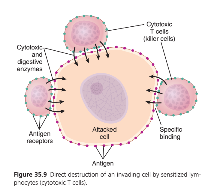
Direct Destruction of an Invading Cell by Cytotoxic T Cells (CD8⁺ Killer T Cells)
This figure explains how Cytotoxic T cells (CD8⁺ T lymphocytes) directly kill virus-infected cells, cancer cells, and transplanted foreign cells.
Unlike B lymphocytes, which kill microorganisms by producing antibodies, cytotoxic T cells destroy abnormal cells directly.
This process is called:
- Cell-mediated cytotoxicity
- Cell-mediated immunity
- Direct killing by Cytotoxic (CD8⁺) T cells
This figure is one of the highest-yield MBBS and USMLE concepts.
Overall Concept
Imagine a city where one house has been occupied by terrorists.
The army cannot destroy the entire city.
Instead,
special commandos surround only that infected house.
They recognize it,
attach to it,
release explosives,
and destroy only that house.
The cytotoxic T cell behaves exactly like these commandos.
- Cytotoxic T cell = Special forces
- Virus-infected cell = Terrorist hideout
- Perforin = Explosive charge
- Granzymes = Assassins entering through the holes
- Cell death = Elimination of the infected target
First Look at the Figure
The figure contains five important components.
- Cytotoxic T cell (killer cell)
- Antigen receptors on the T cell
- Antigen on the attacked cell
- Specific binding between T cell and target cell
- Release of cytotoxic and digestive enzymes
Everything revolves around the interaction between the killer T cell and the infected target cell.
Step-by-Step Sequence
Virus infects body
        ↓
Virus infects a normal body cell
        ↓
Cell displays viral peptide on MHC Class I
        ↓
Cytotoxic T cell recognizes the antigen
        ↓
Specific binding occurs
        ↓
Perforin and granzymes are released
        ↓
Target cell undergoes apoptosis
        ↓
Virus replication stops
PART 1
Cytotoxic T Cell (Killer Cell)
The small pink cells surrounding the large cell represent
Cytotoxic T Cells
Also called
- CD8⁺ T cells
- Killer T cells
- Cytotoxic lymphocytes (CTLs)
Main Function
They destroy
- Virus-infected cells
- Cancer cells
- Transplanted foreign cells
- Cells infected with intracellular bacteria (in some infections)
Unlike antibodies,
they do not attack free microorganisms.
They destroy the infected body cell itself.
Easy Analogy
Suppose a factory becomes infected with dangerous chemicals.
Instead of removing every chemical one by one,
the entire factory is demolished.
That is what a cytotoxic T cell does.
PART 2
Attacked Cell
The large cell in the center represents
an infected or abnormal body cell.
Examples include
- Virus-infected liver cell
- Virus-infected lung cell
- Cancer cell
- Transplanted kidney cell
Why Is It Attacked?
Because abnormal proteins (antigens) are displayed on its surface.
These are recognized as “non-self.”
PART 3
Antigen on the Target Cell
Notice the small purple structures labeled
Antigen
These represent
viral or abnormal peptide fragments.
Where Do These Antigens Come From?
Example:
A virus enters a cell.
↓
The virus produces proteins.
↓
The infected cell breaks some viral proteins into peptides.
↓
These peptides are displayed on
MHC Class I molecules
on the cell surface.
Important Point
All nucleated cells express MHC Class I.
This allows cytotoxic T cells to inspect them for infection.
PART 4
Antigen Receptors
The green dots on the cytotoxic T cell represent
T-Cell Receptors (TCRs)
Function
Each TCR recognizes
only one specific antigen
presented on
MHC Class I.
Lock-and-Key Analogy
TCR = Lock
Antigen + MHC I = Key
Only the correct combination activates the cytotoxic T cell.
PART 5
Specific Binding
The figure labels
Specific Binding
What Happens?
The cytotoxic T cell firmly attaches to the infected cell.
This contact forms an
Immunological Synapse
At this point,
the cytotoxic T cell is ready to kill.
Why Is Specific Binding Important?
Without specific recognition,
healthy cells would also be destroyed.
Specificity ensures
only abnormal cells are targeted.
Easy Analogy
A police officer arrests only the criminal after checking the identity card.
Healthy citizens are left untouched.
PART 6
Release of Cytotoxic and Digestive Enzymes
The arrows entering the attacked cell represent
cytotoxic molecules released by the killer T cell.
The figure labels them as
Cytotoxic and Digestive Enzymes
Modern immunology identifies the main molecules as:
1. Perforin
Perforin forms pores in the target cell membrane.
Think of perforin as a drill making holes in a wall.
2. Granzymes
Granzymes enter through these pores.
They activate enzymes inside the target cell,
leading to
Apoptosis (programmed cell death).Easy Analogy
Imagine burglars drilling a hole in a safe (perforin),
then sending specialists inside to destroy the contents (granzymes).
How Does the Target Cell Die?
The infected cell usually dies by
Apoptosis
rather than bursting immediately.
Why Apoptosis?
Apoptosis is a clean form of cell death.
It minimizes inflammation and prevents unnecessary damage to surrounding tissues.
What Happens After Killing?
The cytotoxic T cell does not die.
Instead,
it detaches,
searches for another infected cell,
and repeats the process.
One cytotoxic T cell can destroy
many infected cells.
Easy Analogy
A trained soldier completes one mission,
then moves on to the next target.Difference Between Cytotoxic T Cells and Natural Killer (NK) Cells
Many students confuse these cells.
| Cytotoxic T Cell (CD8⁺) | Natural Killer (NK) Cell |
|---|---|
| Adaptive immunity | Innate immunity |
| Requires prior sensitization | Acts without prior sensitization |
| Recognizes specific antigen on MHC I | Detects cells with reduced or absent MHC I |
| Has T-cell receptor | No antigen-specific TCR |
Difference Between Cytotoxic T Cells and Antibodies
| Cytotoxic T Cells | Antibodies |
|---|---|
| Kill infected body cells | Neutralize extracellular pathogens and toxins |
| Cell-mediated immunity | Humoral immunity |
| Require MHC I presentation | Bind free antigens directly |
| Use perforin and granzymes | Use neutralization, opsonization, complement activation |
Clinical Correlations
1. Viral Infection
In influenza or hepatitis B infection,
infected cells display viral peptides on MHC I.
Cytotoxic T cells recognize these cells and induce apoptosis, limiting viral spread.
2. Cancer Immunity
Many tumor cells express abnormal antigens.
Cytotoxic T cells recognize these antigens and destroy cancer cells.
This principle is used in modern cancer immunotherapy.
3. Organ Transplant Rejection
Transplanted organs express foreign HLA (MHC) molecules.
Recipient cytotoxic T cells recognize these as foreign and attack the graft unless immunosuppressive drugs are used.
4. HIV Infection
HIV primarily infects CD4⁺ helper T cells.
Without helper T-cell cytokines,
cytotoxic T-cell activation becomes much less effective,
leading to impaired control of viral infections.
Relationship with Previous Figures
| Figure | Main Concept |
|---|---|
| 35.1 | Development of T and B lymphocytes |
| 35.2 | Clonal selection of B cells |
| 35.3 | Primary and secondary antibody responses |
| 35.4 | Structure of antibodies |
| 35.5 | Agglutination by antibodies |
| 35.6 | Complement-mediated destruction |
| 35.8 | Helper T cells coordinate the immune response |
| 35.9 | Cytotoxic T cells directly kill infected or abnormal cells |
Notice the sequence:
- Figure 35.8 shows helper T cells activating cytotoxic T cells through cytokines.
- Figure 35.9 shows what activated cytotoxic T cells actually do—they recognize infected cells and eliminate them.
High-Yield MBBS Points
- Cytotoxic T cells (CD8⁺) are the principal effector cells of cell-mediated immunity.
- They recognize specific peptide antigens presented on MHC Class I molecules.
- All nucleated cells express MHC Class I, allowing surveillance for intracellular infection.
- The T-cell receptor (TCR) binds specifically to the peptide–MHC I complex.
- After specific binding, cytotoxic T cells release:
- Perforin → forms pores in the target-cell membrane.
- Granzymes → enter through the pores and trigger apoptosis.
- One cytotoxic T cell can kill multiple target cells sequentially.
- Cytotoxic T cells are especially important in viral infections, tumor surveillance, and transplant rejection.
Complete Flow Chart
Virus infects a body cell
          ↓
Viral proteins are processed
          ↓
Viral peptide + MHC Class I displayed
          ↓
Recognition by CD8⁺ Cytotoxic T Cell (TCR)
          ↓
Specific Binding (Immunological Synapse)
          ↓
Release of Perforin
          ↓
Pore Formation
          ↓
Entry of Granzymes
          ↓
Activation of Apoptosis
          ↓
Death of the Infected Cell
          ↓
Cytotoxic T Cell Detaches
          ↓
Kills Another Infected Cell
Key Concept
Figure 35.9 illustrates the final effector phase of cell-mediated immunity. Virus-infected or abnormal cells display antigenic peptides on MHC Class I molecules. Sensitized CD8⁺ cytotoxic T cells recognize these peptide–MHC I complexes through their T-cell receptors, bind specifically to the target cell, and release perforin and granzymes. Perforin creates membrane pores, while granzymes trigger apoptosis, leading to precise elimination of infected or malignant cells with minimal damage to surrounding healthy tissue. This mechanism is essential for defense against intracellular pathogens, cancer cells, and transplanted foreign tissues.
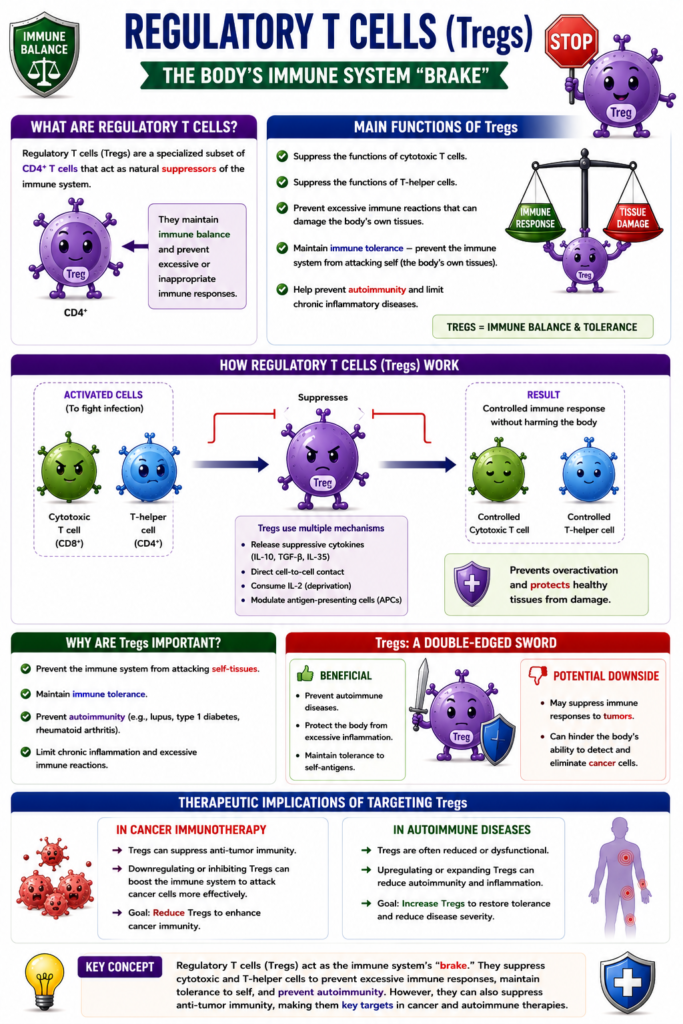
Regulatory T Cells
- Regulatory T cells (Tregs) can suppress the functions of:
- Cytotoxic T cells
- T-helper cells
- These suppressor functions are carried out by CD4⁺ regulatory T cells (Tregs).
- Tregs prevent cytotoxic T cells from producing excessive immune reactions.
- This helps protect the body’s own tissues from damage.
- The regulatory T-cell system plays an important role in immune tolerance.
- Immune tolerance means limiting the immune system’s ability to attack the body’s own tissues.
- Tregs help prevent autoimmunity.
- They also help limit chronic inflammatory diseases.
- However, Tregs may also suppress immune responses against tumors.
- This can reduce the body’s natural ability to control the growth of cancer cells.
- Because of this, much research is focused on immunotherapy.
- In cancer, immunotherapy aims to decrease (downregulate) Treg activity.
- In autoimmune disorders, immunotherapy aims to increase (upregulate) Treg activity.
Key Concept
Regulatory T cells (CD4⁺ Tregs) suppress the activity of cytotoxic and T-helper cells, maintaining immune tolerance and preventing excessive immune reactions, autoimmunity, and chronic inflammation. However, they can also reduce anti-tumor immunity, making Tregs important targets for immunotherapy in both cancer and autoimmune diseases.
MADE BY SELF LEARNING CEO AND FOUNDER DR SHEEN
