


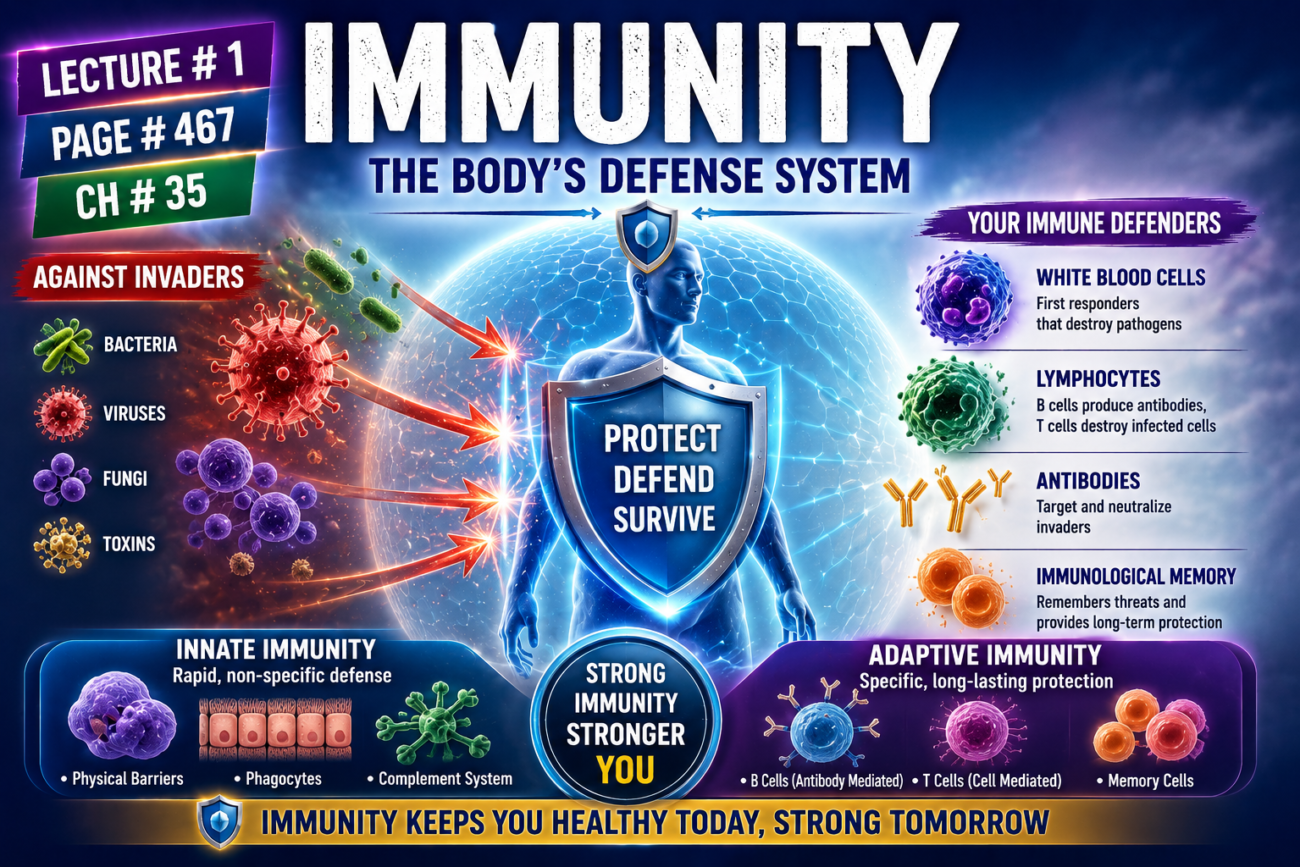
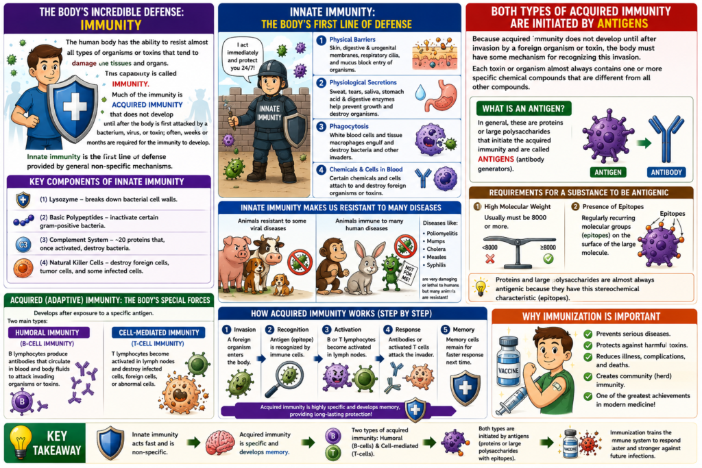
The human body can protect itself from almost all organisms and toxins that can damage tissues and organs.
- This protective ability is called immunity.
- Much of the body’s protection is acquired immunity.
- Acquired immunity develops only after the body is attacked for the first time by a bacterium, virus, or toxin.
- It may take weeks or even months for acquired immunity to fully develop.
- Another type of protection is called innate immunity.
- Innate immunity works through general defense mechanisms.
- It is not directed against one specific disease organism.
- Innate immunity includes barriers that stop harmful substances from entering the body.
- It also provides the first line of defense against invading organisms.
- Physical barriers include:
- Skin
- Digestive tract and urogenital membranes
- Ciliated epithelium of the respiratory tract
- Mucus
- These barriers help prevent organisms from entering the body.
- Physiological secretions include:
- Sweat
- Tears
- Saliva
- These secretions help stop microorganisms from growing.
- Stomach acid and digestive enzymes destroy many organisms that are swallowed.
- White blood cells and tissue macrophages destroy bacteria and other invading organisms by phagocytosis.
- Certain chemicals and cells present in the blood attach to foreign organisms or toxins and destroy them.
- Some important components of innate immunity are:
- Lysozyme: A mucolytic polysaccharide that breaks down bacteria by causing their lysis.
- Basic polypeptides: They react with and inactivate certain gram-positive bacteria.
- Complement complex: About 20 proteins that become activated in different ways to destroy bacteria.
- Natural killer lymphocytes: They recognize and destroy foreign cells, tumor cells, and some infected cells.
- Innate immunity naturally protects humans from some diseases that affect animals.
- Examples include:
- Hog cholera
- Cattle plague
- Distemper, a viral disease that kills many infected dogs.
- In the same way, many animals are naturally resistant or immune to several human diseases.
- Examples include:
- Poliomyelitis
- Mumps
- Human cholera
- Measles
- Syphilis
- These diseases can be very harmful or even fatal in humans.
Key Concept
Immunity protects the body from harmful organisms and toxins. Acquired immunity develops after exposure to a specific pathogen, whereas innate immunity provides immediate, general protection through physical barriers, protective secretions, phagocytic cells, blood chemicals, complement proteins, lysozyme, basic polypeptides, and natural killer lymphocytes.
Acquired (Adaptive) Immunity
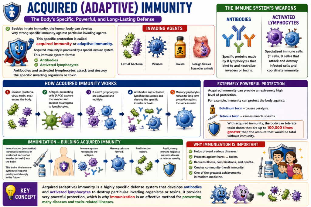
- Besides innate immunity, the human body can develop very strong specific immunity against particular invading agents.
- These invading agents include:
- Lethal bacteria
- Viruses
- Toxins
- Foreign tissues from other animals
- This specific protection is called acquired immunity or adaptive immunity.
- Acquired immunity is produced by a special immune system.
- This immune system forms:
- Antibodies
- Activated lymphocytes
- Antibodies and activated lymphocytes attack and destroy the specific invading organism or toxin.
- Acquired immunity can provide an extremely high level of protection.
- For example, immunity can protect the body against:
- Botulinum toxin, which causes paralysis.
- Tetanus toxin, which causes muscle spasms.
- With acquired immunity, the body can tolerate toxin doses that are up to 100,000 times greater than the amount that would be fatal without immunity.
- Because acquired immunity gives such strong protection, immunization is very important.
- Immunization helps protect people against diseases and harmful toxins.
Key Concept
Acquired (adaptive) immunity is a highly specific defense system that develops antibodies and activated lymphocytes to destroy particular invading organisms or toxins. It provides very powerful protection, which is why immunization is an effective method for preventing many diseases and toxin-related illnesses.
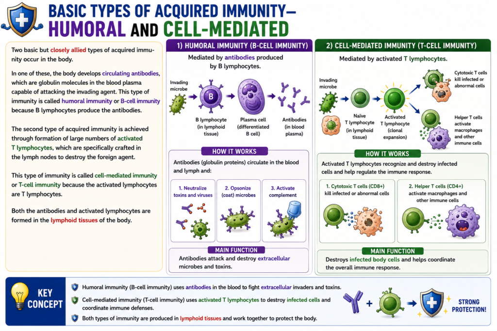
Basic Types of Acquired Immunity—Humoral and Cell-Mediated
- There are two basic and closely related types of acquired immunity in the body.
- The first type is humoral immunity.
- In humoral immunity, the body produces circulating antibodies.
- Antibodies are globulin molecules present in the blood plasma.
- These antibodies attack the invading organism or foreign agent.
- Humoral immunity is also called B-cell immunity.
- It is called B-cell immunity because B lymphocytes produce antibodies.
- The second type is cell-mediated immunity.
- In cell-mediated immunity, the body produces large numbers of activated T lymphocytes.
- These activated T lymphocytes are specially formed in the lymph nodes.
- Their function is to destroy the specific foreign agent.
- Cell-mediated immunity is also called T-cell immunity.
- It is called T-cell immunity because the activated lymphocytes are T lymphocytes.
- Both antibodies and activated lymphocytes are produced in the lymphoid tissues of the body.
Key Concept
Acquired immunity has two main types: humoral (B-cell) immunity, in which B lymphocytes produce antibodies to attack foreign agents, and cell-mediated (T-cell) immunity, in which activated T lymphocytes directly destroy specific invading agents. Both are formed in the body’s lymphoid tissues.
Both Types of Acquired Immunity Are Initiated by Antigens
- Acquired immunity develops only after the body is invaded by a foreign organism or toxin.
- Therefore, the body must have a way to recognize the invading substance.
- Every organism or toxin usually contains one or more unique chemical compounds.
- These chemical compounds are different from the compounds found in other organisms or toxins.
- Most of these unique compounds are:
- Proteins
- Large polysaccharides
- These substances start the process of acquired immunity.
- They are called antigens.
- Antigen means antibody generator.
- For a substance to act as an antigen, it usually must have a high molecular weight of 8000 or more.
- Antigenicity usually depends on regularly repeated molecular groups on the surface of the molecule.
- These repeating molecular groups are called epitopes.
- Epitopes help the immune system recognize the antigen.
- Proteins are almost always antigenic because they contain these repeating stereochemical structures.
- Large polysaccharides are also almost always antigenic for the same reason.
Key Concept
Both humoral and cell-mediated immunity begin when the immune system recognizes antigens. Antigens are usually proteins or large polysaccharides with a molecular weight of 8000 or more. Their repeating surface structures, called epitopes, allow the immune system to recognize them and initiate acquired immunity.
Lymphocytes Are Responsible for Acquired Immunity
- Acquired immunity is produced by the body’s lymphocytes.
- If a person is born with a genetic absence of lymphocytes, acquired immunity cannot develop.
- If lymphocytes are destroyed by radiation or chemicals, acquired immunity also cannot develop.
- A newborn without lymphocytes can die from severe bacterial infection within a few days after birth unless treated with heroic measures.
- Therefore, lymphocytes are essential for human survival.
- Lymphocytes are found mainly in the lymph nodes.
- They are also present in special lymphoid tissues, including:
- Spleen
- Submucosal areas of the gastrointestinal tract
- Thymus
- Bone marrow
- Lymphoid tissues are strategically distributed throughout the body.
- Their location helps them intercept invading organisms and toxins before they spread widely.
- In most cases, an invading organism first enters the tissue fluid.
- It is then carried through the lymph vessels to a lymph node or another lymphoid tissue.
- The lymphoid tissue in the gastrointestinal wall is exposed immediately to antigens entering from the gut.
- The lymphoid tissue of the throat and pharynx, including the tonsils and adenoids, intercepts antigens entering through the upper respiratory tract.
- The lymphoid tissue in the lymph nodes is exposed to antigens that invade the body’s peripheral tissues.
- The spleen, thymus, and bone marrow intercept antigenic agents that have entered the circulating blood.
Key Concept
Lymphocytes are the cells responsible for acquired immunity and are essential for survival. They are concentrated in lymph nodes and other lymphoid tissues, where they intercept antigens entering through tissues, the gastrointestinal tract, the respiratory tract, and the bloodstream before the invaders can spread throughout the body.
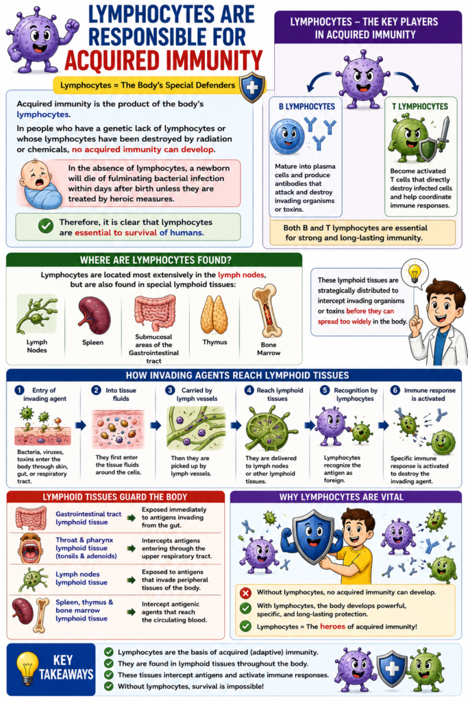
T and B Lymphocytes Promote Cell-Mediated and Humoral Immunity
- Most lymphocytes look similar under a microscope.
- However, lymphocytes are divided into two major populations.
- The first population is T lymphocytes (T cells).
- T lymphocytes produce activated lymphocytes.
- These activated lymphocytes provide cell-mediated immunity.
- The second population is B lymphocytes (B cells).
- B lymphocytes produce antibodies.
- These antibodies provide humoral immunity.
- Both T and B lymphocytes develop from multipotent hematopoietic stem cells in the embryo.
- These stem cells form common lymphoid progenitor cells during differentiation.
- Almost all newly formed lymphocytes eventually move to the lymphoid tissues.
- Before reaching the lymphoid tissues, they undergo further differentiation (preprocessing).
- Lymphoid progenitor cells that will become T lymphocytes first migrate to the thymus gland.
- They are preprocessed in the thymus.
- They are called T lymphocytes because of their association with the thymus.
- T lymphocytes are responsible for cell-mediated immunity.
- Lymphoid progenitor cells that will become B lymphocytes are preprocessed:
- In the liver during mid-fetal life
- In the bone marrow during late fetal life and after birth
- B lymphocytes were first discovered in birds.
- Birds have a special preprocessing organ called the bursa of Fabricius.
- B lymphocytes are named after the bursa of Fabricius.
- B lymphocytes are responsible for humoral immunity by producing antibodies.
Figure Number
- Figure 35.1: Shows the two lymphocyte systems responsible for:
- Formation of activated T lymphocytes
- Formation of antibodies
Key Concept
Lymphocytes are of two main types: T lymphocytes, which mature in the thymus and provide cell-mediated immunity, and B lymphocytes, which mature in the liver (during fetal life) and bone marrow (late fetal life and after birth) and produce antibodies for humoral immunity. Both originate from common lymphoid progenitor cells derived from multipotent hematopoietic stem cells.
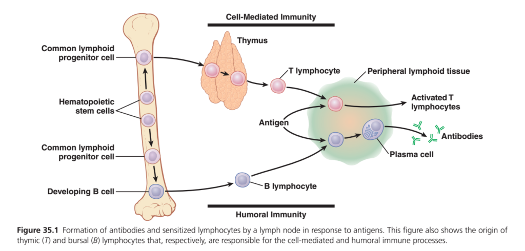
Guyton Physiology Figure 35.1 Explained in Detail
Formation of T Lymphocytes and B Lymphocytes (Cell-Mediated and Humoral Immunity)
This figure is one of the most important diagrams in immunology because it explains:
- Where lymphocytes come from
- How T cells and B cells develop
- Where they mature
- How they become activated
- How antibodies are produced
- Difference between Cell-Mediated Immunity and Humoral Immunity
This single figure summarizes almost the entire adaptive (acquired) immune response.
Overall Concept
Imagine a country preparing for war.
The body also has two different armies.
Army 1
Special commandos
They fight the enemy directly.
These are
T lymphocytes
↓
Called
Cell-Mediated Immunity
Army 2
Weapon manufacturers
Instead of fighting themselves,
they manufacture missiles.
These missiles are
Antibodies
These are produced by
B lymphocytes
↓
Called
Humoral Immunity
So the body fights infection in two different ways.
First Look at the Figure
The figure starts on the left side inside the bone marrow and ends on the right side inside the lymph node (peripheral lymphoid tissue).
The sequence is
Bone marrow
       ↓
Stem cell
       ↓
Common lymphoid progenitor
       ↓
Two pathways

T cell pathway
       ↓
Thymus
       ↓
Mature T lymphocyte
       ↓
Activated T cell

B cell pathway
       ↓
Bone marrow maturation
       ↓
Mature B lymphocyte
       ↓
Plasma cell
       ↓
Antibodies
PART 1
Bone Marrow
The long bone shown on the left represents
Bone Marrow
Bone marrow is called
The Birthplace of Blood Cells
Every blood cell originates here.
Bone marrow produces
- RBCs
- Platelets
- WBCs
- Lymphocytes
Easy Analogy
Think of bone marrow as
A medical university.
Every immune cell begins its life here as a student.
Later,
different students specialize in different fields.
PART 2
Hematopoietic Stem Cells
Inside the bone marrow are
Hematopoietic Stem Cells (HSCs)
These are the mother cells of all blood cells.
Why are they called Stem Cells?
Because one stem cell can produce
- RBC
- Platelets
- Neutrophils
- Monocytes
- Eosinophils
- Basophils
- T cells
- B cells
They have two unique properties:
- Self-renewal – they can make identical stem cells.
- Differentiation – they can develop into specialized blood cells.
Clinical Correlation
In bone marrow transplantation, hematopoietic stem cells from a healthy donor repopulate the recipient’s marrow and restore normal blood cell production.
PART 3
Common Lymphoid Progenitor Cell
The stem cell differentiates into a
Common Lymphoid Progenitor (CLP)
This cell is committed to the lymphocyte lineage.
It can no longer become an RBC or neutrophil.
Instead, it can produce
- T lymphocytes
- B lymphocytes
- Natural Killer (NK) cells (not shown in this figure)
Analogy
Imagine a medical student deciding to specialize in medicine instead of engineering. Once this choice is made, the future career is directed toward immune cells.
PART 4
T-Lymphocyte Pathway (Upper Pathway)
The upper pathway represents
Cell-Mediated Immunity
The CLP leaves the bone marrow and travels to the thymus.
Thymus
The thymus is the primary lymphoid organ where T lymphocytes mature.
“T” stands for Thymus.
The thymus does not produce T cells; it matures them.
What Happens in the Thymus?
Immature T cells undergo:
- Development of T-cell receptors (TCRs)
- Positive selection (recognize self-MHC)
- Negative selection (remove strongly self-reactive cells)
Only a small percentage survive this “training.”
Military Academy Analogy
The thymus is like a military academy.
Cadets are rigorously trained.
Only those who pass the training become elite soldiers.
Mature T Lymphocyte
After leaving the thymus,
the T cell is
- mature
- immunocompetent
- still naïve (it has not yet encountered its specific antigen)
It enters the bloodstream and travels to secondary lymphoid organs.
Peripheral Lymphoid Tissue
The green area represents
Peripheral (Secondary) Lymphoid Tissue
Examples include:
- Lymph nodes
- Spleen
- Tonsils
- Peyer’s patches
- Mucosa-associated lymphoid tissue (MALT)
This is where naïve lymphocytes meet antigens.
Antigen
The figure shows an
Antigen
An antigen is any substance recognized as foreign by the immune system.
Examples:
- Bacteria
- Viruses
- Fungi
- Parasites
- Foreign proteins
- Toxins
Important Point
Antigens do not activate every lymphocyte.
Each lymphocyte recognizes only one specific antigen because it carries a unique receptor.
Activated T Lymphocytes
When a T cell recognizes its specific antigen (usually presented by an antigen-presenting cell via MHC molecules), it becomes activated.
Activated T cells proliferate and differentiate into:
- Helper T cells (CD4⁺) – coordinate the immune response by secreting cytokines.
- Cytotoxic T cells (CD8⁺) – directly kill infected or abnormal cells.
- Regulatory T cells – suppress excessive immune responses.
- Memory T cells – provide long-term immunity.
Clinical Example
A virus infects a liver cell.
Viral peptides are presented on MHC I.
A cytotoxic T cell recognizes the infected cell and destroys it.
This is cell-mediated immunity.
Why Is It Called Cell-Mediated Immunity?
Because cells themselves perform the immune function.
No antibodies are required.
The T cells directly coordinate or kill target cells.
PART 5
B-Lymphocyte Pathway (Lower Pathway)
The lower pathway represents
Humoral Immunity
The common lymphoid progenitor remains in the bone marrow, where it develops into a B lymphocyte.
(In birds, B cells mature in the Bursa of Fabricius, which is why they are called “B” cells. In humans, they mature in the bone marrow.)
Mature B Lymphocyte
The mature B cell enters the bloodstream and migrates to peripheral lymphoid tissues.
Like T cells, it is immunocompetent but naïve until it encounters its specific antigen.
Antigen Recognition by B Cells
When the appropriate antigen binds to the B-cell receptor (BCR), the B cell becomes activated.
Most protein antigens require help from helper T cells for full activation.
Plasma Cell
Activated B cells differentiate into
Plasma Cells
Plasma cells are antibody-producing factories.
They have:
- abundant rough endoplasmic reticulum
- large Golgi apparatus
- very high rates of protein synthesis
Their primary function is to secrete antibodies.
Factory Analogy
A B cell is like a trained engineer.
A plasma cell is the fully operational factory producing millions of antibody molecules.
Antibodies
The green Y-shaped structures represent
Antibodies (Immunoglobulins)
Functions include:
- Neutralizing viruses and toxins
- Opsonizing bacteria for phagocytosis
- Activating the complement system
- Preventing microbial attachment to host cells
Major antibody classes:
- IgG
- IgA
- IgM
- IgE
- IgD
Clinical Example
Following hepatitis B vaccination:
- The vaccine antigen activates B cells.
- Plasma cells produce anti-hepatitis B antibodies.
- Memory B cells remain for rapid protection against future exposure.
Cell-Mediated vs Humoral Immunity
| Feature | Cell-Mediated Immunity | Humoral Immunity |
|---|---|---|
| Main cell | T lymphocyte | B lymphocyte |
| Maturation site | Thymus | Bone marrow |
| Main effector | Activated T cells | Plasma cells and antibodies |
| Acts against | Virus-infected cells, intracellular pathogens, tumors | Extracellular bacteria, toxins, many viruses before cell entry |
| Antibodies involved? | No | Yes |
Primary vs Secondary Lymphoid Organs
| Primary Lymphoid Organs | Secondary Lymphoid Organs |
|---|---|
| Bone marrow | Lymph nodes |
| Thymus | Spleen |
| Site of lymphocyte development and maturation | Site where lymphocytes encounter antigens and become activated |
Clinical Correlations
1. DiGeorge Syndrome
Failure of thymic development leads to deficient T-cell maturation, causing impaired cell-mediated immunity and recurrent viral and fungal infections.
2. X-linked Agammaglobulinemia (Bruton Disease)
B cells fail to mature, resulting in markedly reduced antibody production and recurrent bacterial infections.
3. HIV Infection
HIV primarily infects CD4⁺ helper T cells, weakening both cell-mediated immunity and the helper signals required for optimal antibody production.
4. Vaccination
Vaccines activate both B and T lymphocytes, generating memory cells that provide faster and stronger immune responses upon future exposure.
High-Yield MBBS Points
- All lymphocytes originate from hematopoietic stem cells in the bone marrow.
- Common lymphoid progenitor cells give rise to T cells, B cells, and NK cells.
- T lymphocytes mature in the thymus and mediate cell-mediated immunity.
- B lymphocytes mature in the bone marrow and mediate humoral immunity.
- Peripheral lymphoid tissues are the sites where mature naïve lymphocytes encounter antigens.
- Activated B cells differentiate into plasma cells, which secrete antibodies.
- Activated T cells coordinate immune responses, kill infected cells, regulate immunity, and form memory cells.
- Memory B and T cells are responsible for long-lasting immunity following infection or vaccination.
Complete Flow Chart
Hematopoietic stem cell (Bone marrow)
               ↓
Common lymphoid progenitor
               ↓
      ┌──────────────────────────┐
      │                          │
      ▼                          ▼
T-cell lineage              B-cell lineage
      │                          │
Maturation in Thymus      Maturation in Bone marrow
      │                          │
Naïve T cell              Naïve B cell
      │                          │
Migration to secondary lymphoid organs
      │                          │
Encounter with specific antigen
      │                          │
Activated T cells         Plasma cells + Memory B cells
      │                          │
Cell-mediated immunity    Antibody production (Humoral immunity)
Key Concept
Figure 35.1 illustrates the complete developmental pathway of adaptive immunity. All lymphocytes originate from hematopoietic stem cells in the bone marrow. T lymphocytes mature in the thymus and provide cell-mediated immunity by directly coordinating or destroying infected cells. B lymphocytes mature in the bone marrow and provide humoral immunity by differentiating into plasma cells that secrete antibodies. Both cell types become activated only after encountering their specific antigen in peripheral lymphoid organs, ensuring a highly specific and long-lasting immune response.
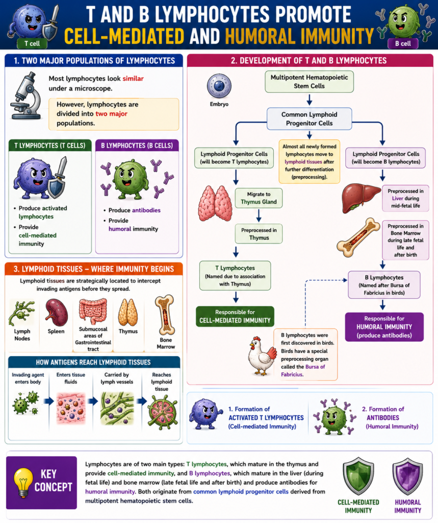
Development and Maturation of T and B Lymphocytes
- All lymphocytes in the body originate from lymphocyte-committed stem cells in the embryo.
- These stem cells cannot directly produce activated T lymphocytes or antibodies.
- Before becoming functional, they must undergo further differentiation (preprocessing) in specific processing areas.
- T lymphocytes are first preprocessed in the thymus gland.
- After originating in the bone marrow, T lymphocytes migrate to the thymus.
- In the thymus, they divide rapidly.
- During this process, they develop extensive diversity to recognize different specific antigens.
- One thymic lymphocyte becomes specific for one antigen.
- The next thymic lymphocyte becomes specific for another antigen.
- This process continues until thousands of different types of T lymphocytes are formed.
- These T lymphocytes can recognize many thousands of different antigens.
- After preprocessing, the mature T lymphocytes leave the thymus.
- They travel through the bloodstream.
- They settle in lymphoid tissues throughout the body.
- The thymus also prevents T lymphocytes from attacking the body’s own tissues.
- It does this by exposing developing T lymphocytes to the body’s own (self) antigens.
- If a T lymphocyte reacts against a self-antigen, it is destroyed and phagocytized.
- Up to 90% of developing T lymphocytes are eliminated during this selection process.
- Only T lymphocytes that do not react against self-antigens are released into the circulation.
- These surviving T lymphocytes respond only to foreign antigens, such as:
- Bacteria
- Toxins
- Transplanted tissues from another person
- Most T-lymphocyte preprocessing occurs:
- Shortly before birth
- During the first few months after birth
- Removing the thymus gland after this period reduces, but does not completely eliminate, the T-cell immune system.
- Removing the thymus several months before birth can prevent the development of cell-mediated immunity.
- Without normal T-cell development, the body cannot properly reject transplanted organs.
Key Concept
All lymphocytes originate from embryonic stem cells but require preprocessing before becoming functional. T lymphocytes mature in the thymus, where they develop specificity for thousands of foreign antigens while self-reactive cells are eliminated. This selection process ensures self-tolerance and allows mature T lymphocytes to provide effective cell-mediated immunity against foreign antigens.
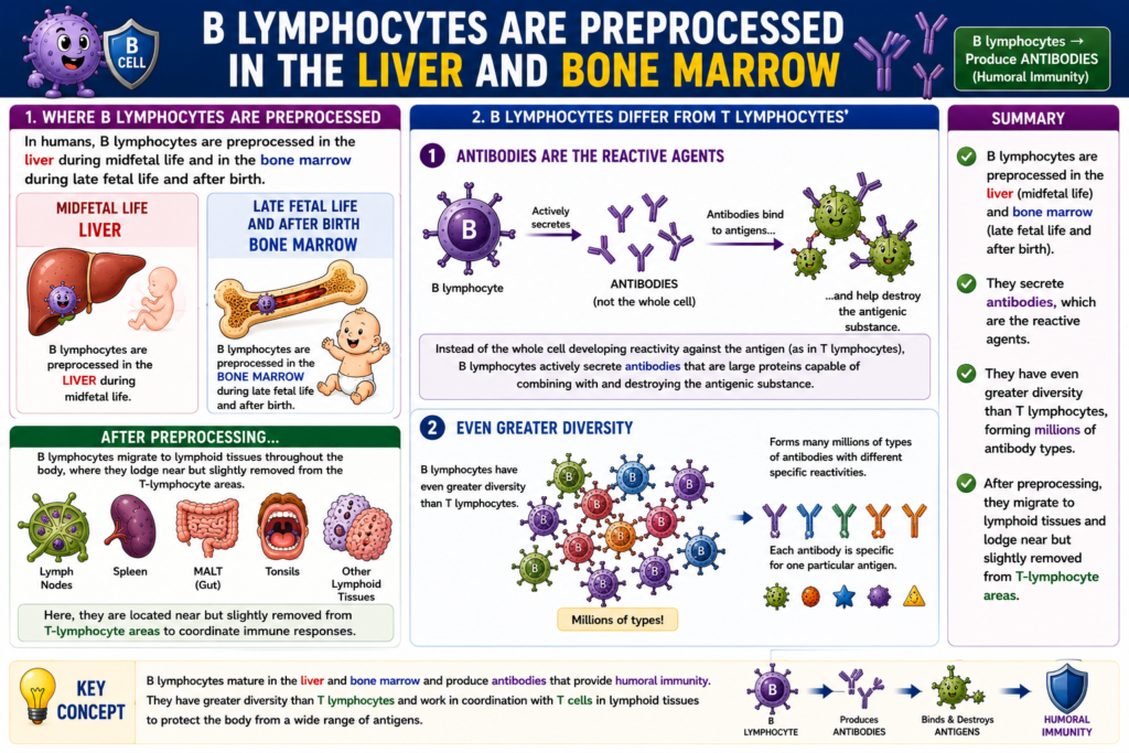
B Lymphocytes Are Preprocessed in the Liver and Bone Marrow
- In humans, B lymphocytes are preprocessed in the liver during mid-fetal life.
- During late fetal life and after birth, they are preprocessed in the bone marrow.
- B lymphocytes differ from T lymphocytes in two important ways.
- Difference 1:
- T lymphocytes themselves become reactive against specific antigens.
- B lymphocytes do not act as the main reactive agents.
- Instead, B lymphocytes actively secrete antibodies.
- Antibodies are large proteins.
- These antibodies combine with the antigen.
- After binding to the antigen, antibodies help destroy the antigenic substance.
- Difference 2:
- B lymphocytes have greater diversity than T lymphocytes.
- They produce many millions of different types of antibodies.
- Each antibody has specific reactivity against a particular antigen.
- After preprocessing, B lymphocytes migrate throughout the body.
- They travel to the lymphoid tissues, just like T lymphocytes.
- In the lymphoid tissues, B lymphocytes are located near, but slightly separated from, the T-lymphocyte areas.
Key Concept
B lymphocytes mature in the liver during mid-fetal life and in the bone marrow during late fetal life and after birth. Unlike T lymphocytes, B lymphocytes produce antibodies that destroy antigens. They also have greater diversity, allowing the formation of millions of different antibodies with specific antigen recognition, and they later migrate to lymphoid tissues near T-cell areas.
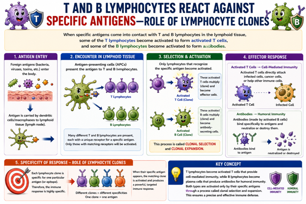
T Lymphocytes and B-Lymphocyte Antibodies React Against Specific Antigens—Role of Lymphocyte Clones
- Specific antigens come into contact with T lymphocytes and B lymphocytes in the lymphoid tissues.
- Some T lymphocytes become activated after recognizing a specific antigen.
- These activated T lymphocytes form activated T cells.
- Some B lymphocytes also become activated after recognizing a specific antigen.
- Activated B lymphocytes produce antibodies.
- The activated T cells react only against the specific antigen that activated them.
- The antibodies also react only against the specific antigen that stimulated their formation.
- Therefore, both activated T cells and antibodies are highly specific for the antigen that initiated their development.
Key Concept
When a specific antigen enters the body, it activates matching T and B lymphocytes in the lymphoid tissues. Activated T cells and antibodies then respond only to that particular antigen, providing highly specific acquired immunity.
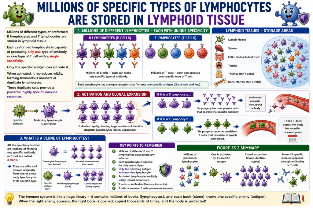
Millions of Specific Types of Lymphocytes Are Stored in Lymphoid Tissue
- The lymphoid tissues store millions of different types of preformed B lymphocytes.
- These B lymphocytes can produce highly specific antibodies.
- The lymphoid tissues also store preformed T lymphocytes.
- Each preformed lymphocyte can produce only one specific type of antibody or one specific type of T cell.
- Each lymphocyte recognizes only one specific antigen.
- Only its matching antigen can activate that lymphocyte.
- Once activated by its specific antigen, the lymphocyte multiplies rapidly.
- It produces a very large number of identical duplicate lymphocytes.
- If the activated cell is a B lymphocyte:
- Its daughter cells produce the same specific antibody.
- The antibody is released into the blood circulation and travels throughout the body.
- If the activated cell is a T lymphocyte:
- Its daughter cells become specific sensitized T cells.
- These T cells enter the lymph.
- They are carried into the blood.
- They circulate through the tissue fluids.
- They return to the lymphatic system.
- This circulation may continue for months or even years.
- All lymphocytes that produce the same specific antibody or the same specific T cell are called a clone of lymphocytes.
- The lymphocytes in a clone are genetically identical.
- They originate from one or a few original lymphocytes of the same specific type.
Figure Number
- Figure 35.2: Shows rapid multiplication (clonal expansion) of a specific activated lymphocyte to produce many identical lymphocytes.
Key Concept
Lymphoid tissues contain millions of preformed lymphocytes, each specific for one antigen. When the correct antigen activates a lymphocyte, it undergoes rapid clonal expansion. B-cell clones produce large amounts of one specific antibody, whereas T-cell clones produce many identical sensitized T cells that circulate throughout the body to provide specific immune protection.
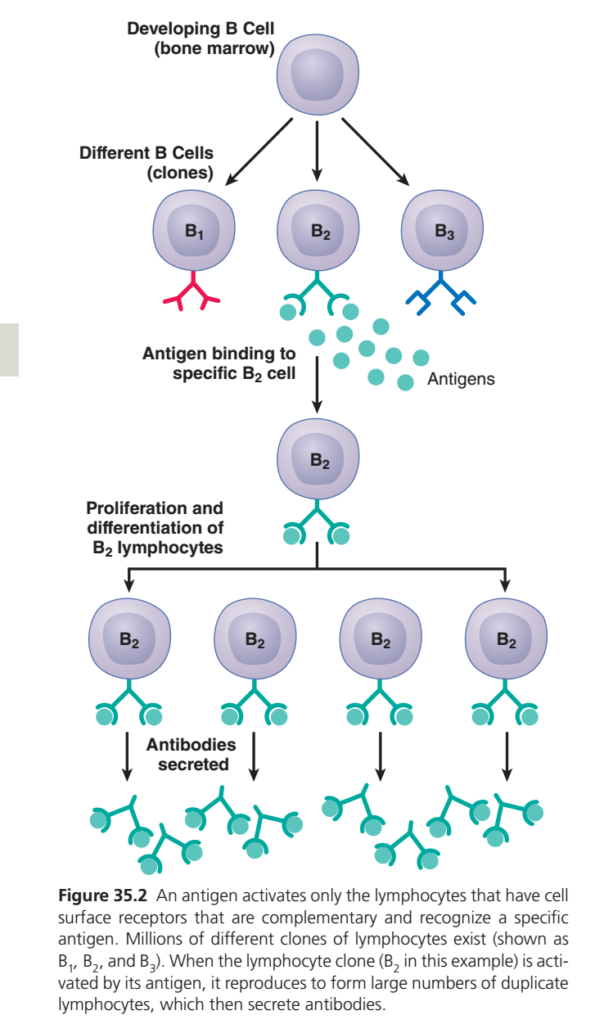
Guyton Physiology Figure 35.2 Explained in Detail
Clonal Selection Theory of B Lymphocytes (How One Antigen Produces Millions of Specific Antibodies)
This figure explains one of the most fundamental concepts of immunology:
How does the body produce an antibody against only one specific antigen while ignoring millions of other substances?
The answer is the Clonal Selection Theory.
This theory explains why our immune system is highly specific and remembers previous infections.
Overall Concept
Imagine a university with 10,000 students.
Each student knows only one foreign language.
| Student | Language |
|---|---|
| Student 1 | English |
| Student 2 | French |
| Student 3 | Japanese |
| Student 4 | Arabic |
| Student 5 | Chinese |
Now suppose a Japanese tourist arrives.
Who can communicate with him?
Only the student who knows Japanese.
That student is selected.
He then trains hundreds of new students who all know Japanese.
Exactly the same thing happens in this figure.
First Look at the Figure
The figure can be divided into five sequential steps:
Developing B cell
        ↓
Formation of millions of different B-cell clones
        ↓
Specific antigen binds to only one clone
        ↓
Selected clone multiplies rapidly
        ↓
Plasma cells produce large amounts of antibodies
Everything begins in the bone marrow.
PART 1
Developing B Cell (Bone Marrow)
Look at the top cell.
It represents a
Developing B Cell
This immature B cell develops inside the bone marrow, where it rearranges its immunoglobulin (Ig) genes to create a unique B-cell receptor (BCR).
What Happens During Development?
Every developing B cell randomly rearranges its antibody genes (V, D, and J gene segments).
As a result:
- Every B cell gets one unique receptor
- No two B cells are exactly alike
- The body generates millions of different B-cell specificities
This process occurs before any infection.
Easy Analogy
Imagine a factory making millions of different locks.
Each lock has a different shape.
Later,
only the correct key can open one particular lock.
PART 2
Different B Cells (Clones)
The figure shows three different B cells:
- B₁
- B₂
- B₃
These represent different clones.
What is a Clone?
A clone is a group of lymphocytes that:
- originated from one parent B cell
- have identical receptors
- recognize the same antigen
Every clone is specific for one antigen only.
Notice the Receptors
Look carefully at the receptors.
Each B cell has a different receptor shape.
- B₁ → Red receptor
- B₂ → Green receptor
- B₃ → Blue receptor
These shapes symbolize different antigen-binding sites.
Why Are the Shapes Different?
Because every B cell recognizes only one antigen.
Think of each receptor as a lock.
Only the correct antigen (the key) fits.
Key Point
Before infection,
the body already contains millions of B-cell clones.
They are waiting silently.
Easy Analogy
Imagine millions of different keys stored in a locker.
When the correct lock appears,
only the matching key is useful.
PART 3
Antigen Binding to Specific B₂ Cell
The green circles represent
Antigens
These are foreign substances such as:
- bacteria
- viruses
- toxins
- pollen
- parasites
What Happens?
The antigen moves through the body.
It encounters:
- B₁ ❌ No match
- B₂ ✅ Perfect match
- B₃ ❌ No match
Only B₂ recognizes it.
Why Only B₂?
Because its receptor is complementary to the antigen.
This is similar to:
- Lock and key
- Puzzle pieces
- Barcode and scanner
Only a perfect fit activates the B cell.
Clinical Example
If the invading organism is the hepatitis B virus, only the B-cell clone with receptors specific for hepatitis B antigens will be activated. The countless other B-cell clones remain inactive.
PART 4
Activation of the B₂ Cell
When the antigen binds to B₂,
the B cell becomes activated.
In most protein antigen responses, this activation is strengthened by helper T cells (CD4⁺) through:
- CD40–CD40L interaction
- Cytokines such as IL-4, IL-5, and IL-21
(These helper T-cell interactions are not shown in this simplified figure.)
What Happens After Activation?
The activated B₂ cell receives the message:
“Multiply rapidly and produce antibodies!”
PART 5
Proliferation (Clonal Expansion)
This is the central concept of the figure.
The activated B₂ cell undergoes repeated mitotic divisions.
One cell becomes:
- 2
- 4
- 8
- 16
- 32
- 64
- Hundreds
- Thousands
- Millions
All of these daughter cells are genetically identical clones.
Why Is It Called Clonal Expansion?
Because:
One selected B cell
↓
Produces a large clone of identical B cells.
Easy Analogy
Suppose one excellent teacher trains 500 new teachers.
All 500 teach the same subject.
Similarly,
one activated B cell produces thousands of identical cells that recognize the same antigen.
PART 6
Differentiation
After proliferation,
the cloned B cells differentiate into two major populations:
1. Plasma Cells
These are antibody-producing factories.
Their main function is to secrete massive amounts of antibodies.
2. Memory B Cells
Although not illustrated in this figure, some activated B cells become memory B cells, which remain in the body for years and respond rapidly if the same antigen enters again.
Plasma Cells
Each plasma cell produces only one type of antibody.
Because all plasma cells originated from the same B₂ clone,
all the antibodies are identical.
Why Is This Important?
Every antibody can bind only to the same antigen that activated the original B₂ cell.
This provides remarkable specificity.
Easy Analogy
Imagine one factory producing only one model of car.
Every car leaving the factory has the same design.
Similarly,
every plasma cell from the same clone produces identical antibodies.
PART 7
Antibodies Secreted
At the bottom of the figure,
the Y-shaped structures represent
Antibodies (Immunoglobulins)
These antibodies circulate in:
- Blood
- Lymph
- Tissue fluids
They bind specifically to the original antigen.
Functions of Antibodies
Once attached to the antigen, antibodies help eliminate it by:
- Neutralizing toxins and viruses
- Blocking microbial attachment to host cells
- Opsonizing microbes for phagocytosis
- Activating the complement system
- Agglutinating microorganisms
Why Aren’t B₁ and B₃ Activated?
This is one of the most important concepts.
B₁ and B₃ remain inactive because:
- Their receptors do not fit the antigen.
- Without receptor binding, there is no activation.
- Therefore, there is no proliferation and no antibody production.
This ensures that the immune response remains highly specific.
Why Is Clonal Selection Important?
Without clonal selection:
- Antibodies would attack many harmless molecules.
- Immune responses would be inefficient.
- Specific immunity and immune memory would not exist.
Clonal selection ensures that:
- Only the correct lymphocyte responds.
- Only the needed antibodies are produced.
- The response becomes stronger as the selected clone expands.
Clinical Correlations
1. Vaccination
A vaccine introduces a harmless form of an antigen.
Specific B-cell clones are selected and activated.
They produce plasma cells and memory B cells.
On future exposure to the real pathogen, memory B cells respond rapidly with a much stronger antibody response.
2. Multiple Myeloma
Multiple myeloma is a cancer of a single plasma-cell clone.
As a result:
- One abnormal clone proliferates uncontrollably.
- Large amounts of a single monoclonal antibody (M protein) are produced.
This disease is a pathological example of clonal expansion.
3. Monoclonal Antibody Therapy
Scientists intentionally isolate one B-cell clone and produce identical antibodies in the laboratory.
These monoclonal antibodies are used to treat:
- Cancers
- Autoimmune diseases
- Certain infections
High-Yield MBBS Points
- Each B lymphocyte expresses only one unique B-cell receptor (BCR).
- Millions of different B-cell clones are generated before antigen exposure through gene rearrangement.
- Only the clone with a complementary receptor is activated by a specific antigen.
- Activation leads to clonal expansion (rapid proliferation of identical cells).
- Activated B cells differentiate into plasma cells (antibody secretion) and memory B cells (long-term immunity).
- All antibodies produced by one clone are identical and recognize the same antigen.
- This principle is called the Clonal Selection Theory, proposed by Sir Frank Macfarlane Burnet.
Complete Flow Chart
Hematopoietic Stem Cell
          ↓
Developing B Cell (Bone Marrow)
          ↓
Formation of millions of different B-cell clones
(B₁, B₂, B₃, ...)
          ↓
Specific antigen enters the body
          ↓
Only the complementary B-cell clone binds the antigen
          ↓
B-cell activation
          ↓
Clonal expansion (rapid mitosis)
          ↓
Differentiation
      ┌───────────────┐
      │               │
      ▼               ▼
 Plasma Cells     Memory B Cells
      │               │
Antibody secretion  Long-term immune memory
Concept Integration with Figures 35.1 and 35.2
- Figure 35.1 explained where B cells come from and how they mature before reaching peripheral lymphoid tissues.
- Figure 35.2 explains what happens after a mature B cell encounters its specific antigen: only the matching clone is selected, undergoes clonal expansion, and differentiates into plasma cells and memory B cells.
- Together, these figures describe the complete pathway of humoral immunity: development → antigen recognition → clonal selection → antibody production → immunological memory.
Key Concept
Figure 35.2 illustrates the Clonal Selection Theory, the cornerstone of adaptive immunity. Millions of unique B-cell clones are generated before birth, each carrying a distinct antigen receptor. When a foreign antigen enters the body, it activates only the B-cell clone with the matching receptor. That selected clone rapidly proliferates (clonal expansion) and differentiates into plasma cells that secrete highly specific antibodies, while some cells become memory B cells. This mechanism explains the extraordinary specificity, efficiency, and long-lasting memory of the humoral immune response.
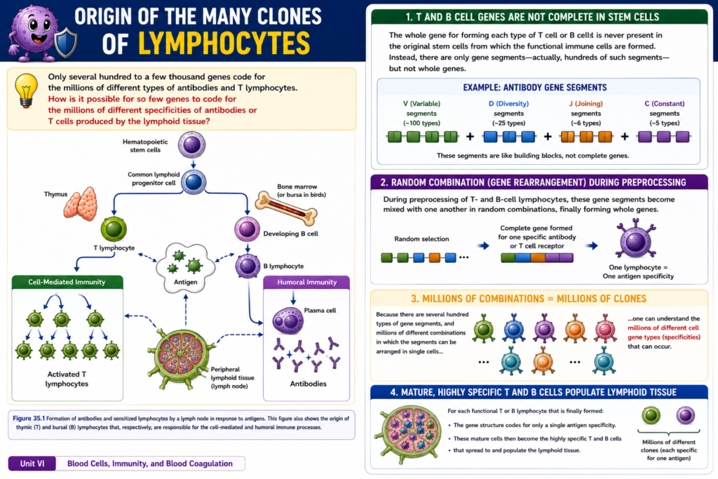
Origin of the Many Clones of Lymphocytes
- Only several hundred to a few thousand genes are available to produce the immune system.
- Yet, the body can produce millions of different types of antibodies and T lymphocytes.
- This is possible because the complete gene is not present in the original stem cells.
- Instead, the original stem cells contain many gene segments.
- There are hundreds of different gene segments.
- During preprocessing of T lymphocytes and B lymphocytes, these gene segments are randomly mixed and combined.
- These random combinations form complete functional genes.
- Because there are hundreds of gene segments, they can be arranged in millions of different combinations.
- As a result, the body can produce millions of different T cells and B cells.
- Each mature T lymphocyte or B lymphocyte contains a gene structure that recognizes only one specific antigen.
- Each mature lymphocyte therefore has only one antigen specificity.
- These highly specific mature T and B lymphocytes spread throughout the lymphoid tissues of the body.
Figure Number
- Figure 35.1: Shows:
- Origin of hematopoietic stem cells
- Formation of common lymphoid progenitor cells
- Development of T lymphocytes in the thymus
- Development of B lymphocytes
- Cell-mediated immunity by activated T lymphocytes
- Humoral immunity by plasma cells producing antibodies
- Response of lymphocytes to an antigen
Key Concept
Millions of different lymphocyte clones are produced through random rearrangement of hundreds of gene segments during T- and B-cell development. Each mature lymphocyte acquires one unique antigen specificity and then populates the lymphoid tissues, allowing the immune system to recognize millions of different foreign antigens.
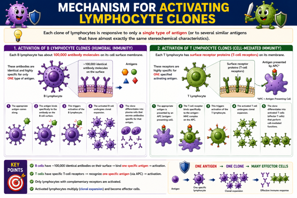
Mechanism for Activating Lymphocyte Clones
- Each clone of lymphocytes responds to only one specific antigen.
- A lymphocyte clone may also respond to a few closely related antigens that have almost the same stereochemical structure.
- In B lymphocytes, the cell surface contains about 100,000 antibody molecules.
- These antibody molecules are present on the cell membrane.
- Each antibody molecule is highly specific for one type of antigen.
- When the matching antigen enters the body, it immediately binds to the antibody on the B-cell membrane.
- This binding activates the B lymphocyte.
- In T lymphocytes, the cell membrane contains surface receptor proteins, also called T-cell receptors (TCRs).
- T-cell receptors are similar to antibodies.
- Each T-cell receptor is highly specific for one activating antigen.
- An antigen activates only those T lymphocytes that have matching (complementary) receptors.
- Therefore, only lymphocytes that are already committed to recognize that antigen become activated.
Key Concept
Each lymphocyte clone is specific for one antigen. B lymphocytes are activated when an antigen binds to the antibody molecules on their surface, while T lymphocytes are activated when an antigen binds to their specific T-cell receptors. Only lymphocytes with complementary receptors respond to a particular antigen, ensuring highly specific acquired immunity.
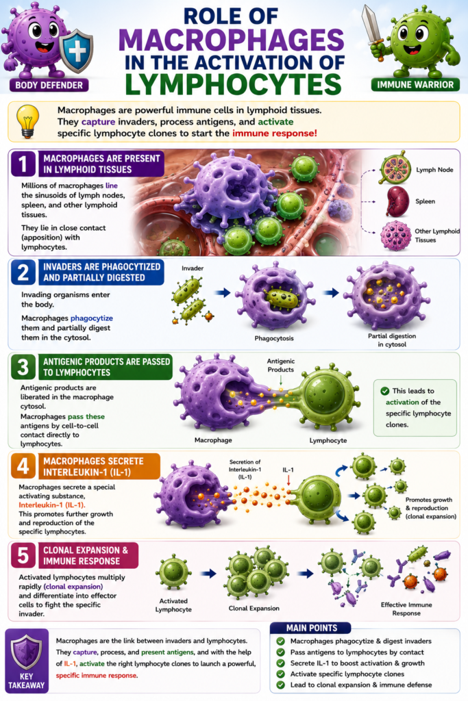
Role of Macrophages in the Activation Process
- In addition to lymphocytes, millions of macrophages are present in the lymphoid tissues.
- These macrophages line the sinusoids of:
- Lymph nodes
- Spleen
- Other lymphoid tissues
- Macrophages are located very close to many lymphocytes in the lymph nodes.
- Most invading organisms are first phagocytized by macrophages.
- Inside the macrophages, the organisms are partially digested.
- During digestion, antigenic products are released into the macrophage cytosol.
- The macrophages then transfer these antigens directly to lymphocytes through cell-to-cell contact.
- This direct transfer activates the specific lymphocyte clones that recognize the antigen.
- Macrophages also secrete a special activating substance called interleukin-1 (IL-1).
- Interleukin-1 further stimulates the growth and multiplication of the specific activated lymphocytes.
Key Concept
Macrophages play a vital role in activating acquired immunity. They phagocytize and partially digest invading organisms, transfer the released antigens directly to specific lymphocytes, and secrete interleukin-1 to promote the growth and proliferation of the activated lymphocyte clones.
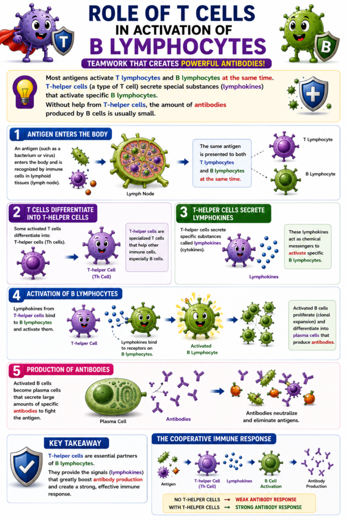
Role of T Cells in Activation of B Lymphocytes
- Most antigens activate both T lymphocytes and B lymphocytes at the same time.
- Some of the activated T lymphocytes become T-helper cells.
- T-helper cells secrete special chemical substances.
- These substances are collectively called lymphokines.
- Lymphokines activate specific B lymphocytes.
- Activated B lymphocytes then produce antibodies.
- Without the help of T-helper cells, B lymphocytes produce only a small amount of antibodies.
- Therefore, T-helper cells are important for a strong antibody response.
- The cooperative relationship between helper T cells and B lymphocytes is explained later after the mechanisms of T-cell immunity are described.
Key Concept
Most antigens activate both T and B lymphocytes simultaneously. T-helper cells release lymphokines that stimulate specific B lymphocytes, greatly increasing antibody production. Without T-helper cell support, the antibody response is usually weak.
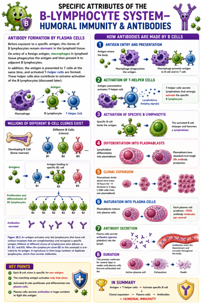
Specific Attributes of the B-Lymphocyte System—Humoral Immunity and Antibodies
- Before exposure to a specific antigen, the B-lymphocyte clones remain dormant in the lymphoid tissues.
- When a foreign antigen enters the body, macrophages in the lymphoid tissues phagocytize the antigen.
- The macrophages then present the antigen to nearby B lymphocytes.
- At the same time, the antigen is also presented to T lymphocytes.
- This leads to the formation of activated T-helper cells.
- T-helper cells further increase the activation of B lymphocytes.
- The B lymphocytes that recognize the specific antigen rapidly enlarge.
- These enlarged B lymphocytes are called lymphoblasts.
- Some lymphoblasts further differentiate into plasmablasts.
- Plasmablasts are the precursors of plasma cells.
- In plasmablasts:
- The cytoplasm enlarges.
- The rough endoplasmic reticulum increases greatly.
- Plasmablasts divide about once every 10 hours.
- They undergo about 9 cell divisions in 4 days.
- This produces about 500 cells from one original plasmablast.
- The mature plasma cells produce gamma globulin antibodies at a very rapid rate.
- Each plasma cell produces about 2000 antibody molecules every second.
- These antibodies are released into the lymph.
- The lymph carries the antibodies into the circulating blood.
- Antibody production continues for several days or weeks.
- Eventually, the plasma cells become exhausted and die.
Figure Number
- Figure 35.2: Shows:
- Different B-lymphocyte clones (B1, B2, B3)
- Binding of an antigen to the specific B-cell clone (B2)
- Clonal proliferation and differentiation of the activated B lymphocyte
- Formation of many identical B lymphocytes
- Secretion of antibodies by the activated clone
Key Concept
B-lymphocyte clones remain inactive until they encounter their specific antigen. After antigen presentation by macrophages and assistance from T-helper cells, the activated B lymphocytes become lymphoblasts, then plasmablasts, and finally plasma cells. Plasma cells rapidly produce and secrete large amounts of gamma globulin antibodies into the lymph and blood until they eventually become exhausted and die.
Formation of Memory Cells Enhances Antibody Response to Subsequent Antigen Exposure
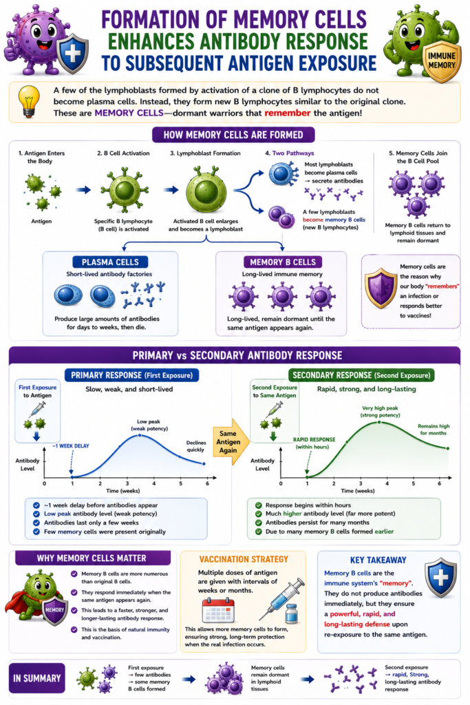
- After activation of a B-lymphocyte clone, some lymphoblasts do not become plasma cells.
- Instead, they develop into new B lymphocytes that are similar to the original clone.
- As a result, the B-cell population of that specific clone increases greatly.
- These new B lymphocytes are added to the original clone.
- They circulate throughout the body.
- They populate the lymphoid tissues.
- These B lymphocytes remain immunologically dormant.
- They become active only when the same antigen enters the body again.
- These long-lived B lymphocytes are called memory cells.
- When the body is exposed to the same antigen again, the memory cells respond.
- The second antibody response is much faster.
- It is also much stronger.
- This occurs because many more memory cells are present than the number of original B lymphocytes.
- The primary antibody response occurs after the first exposure to an antigen.
- The primary response has:
- About a 1-week delay before antibodies appear.
- Weak potency.
- Short duration.
- The secondary antibody response occurs after the second exposure to the same antigen.
- The secondary response:
- Begins rapidly, often within hours.
- Is much more powerful.
- Continues producing antibodies for many months instead of only a few weeks.
- Because the secondary response is stronger and lasts longer, immunization is usually given in multiple doses.
- These doses are separated by several weeks or several months.
Figure Number
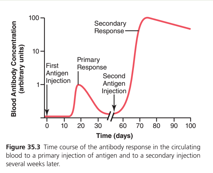
- Figure 35.3: Shows the difference between:
- Primary antibody response after the first exposure to an antigen.
- Secondary antibody response after the second exposure to the same antigen, demonstrating a faster, stronger, and longer-lasting antibody production.
Key Concept
Some activated B lymphocytes become memory cells instead of plasma cells. These memory cells remain dormant until the same antigen is encountered again, producing a rapid, powerful, and long-lasting secondary antibody response. This memory response is the basis for the effectiveness of booster doses during immunization.
Guyton Physiology Figure 35.3 Explained in Detail
Primary and Secondary Immune (Antibody) Response
This figure is one of the highest-yield concepts in immunology because it explains:
- Why the first infection usually causes a slower immune response.
- Why the second exposure to the same organism produces a much faster and stronger response.
- How memory B cells work.
- Why vaccines are effective.
This graph forms the basis of immunological memory, one of the defining features of the adaptive immune system.
Overall Concept
Imagine you are appearing for an MBBS exam.
First Time
You have never studied the subject before.
You need time to:
- read books
- understand concepts
- practice questions
Your performance is initially slow.
Second Time
You appear for the same exam again.
Now you already know:
- the syllabus
- important topics
- common questions
You answer much faster and score much higher.
The immune system behaves in exactly the same way.
First Look at the Graph
The graph has two axes.
X-axis
Time (Days)
This shows how much time passes after antigen exposure.
Y-axis
Blood Antibody Concentration
This shows how many antibodies are present in the blood.
Notice that the scale is logarithmic (1, 10, 100), meaning each step represents a tenfold increase rather than a simple linear increase.
The higher the curve,
the greater the antibody concentration.
Important Events in the Graph
There are four major events:
- First antigen injection
- Primary immune response
- Second antigen injection
- Secondary immune response
Each event tells an important story.
PART 1
First Antigen Injection (Day 0)
Look at the left side of the graph.
At Day 0, the antigen enters the body for the first time.
This may occur through:
- bacterial infection
- viral infection
- vaccination
- tetanus injection
The immune system has never seen this antigen before.
What Happens Immediately?
Surprisingly,
almost nothing happens at first.
The antibody level remains close to zero.
This period is called the
Lag Phase
Why Does the Lag Phase Occur?
Because the immune system must first:
- recognize the antigen
- select the correct B-cell clone (clonal selection)
- activate helper T cells (for most protein antigens)
- stimulate B-cell proliferation
- differentiate B cells into plasma cells
All of these processes take time.
Usually,
the lag phase lasts about 5–10 days, although the exact duration varies with the antigen.
Easy Analogy
Suppose a thief enters a city.
First,
the police must:
- receive the complaint
- identify the thief
- gather officers
- prepare equipment
Only then does the operation begin.
PART 2
Primary Immune Response
Around the second week, antibody production begins.
The curve slowly rises.
This is called the
Primary Immune Response
What Happens?
The activated B cells become plasma cells.
Plasma cells begin secreting antibodies.
Initially,
the main antibody produced is:
IgM
Later,
with class switching,
larger amounts of:
IgG
are produced.
Why Is the Response Small?
Only a few naïve B cells recognize the antigen.
These cells require time for:
- clonal expansion
- plasma-cell differentiation
- antibody secretion
Therefore,
the response is:
- slow
- relatively weak
- short-lived
Clinical Example
A person becomes infected with the hepatitis A virus for the first time.
The body requires several days before producing detectable antibodies.
During this period,
the person develops symptoms.
Why Does the Curve Fall Again?
After the infection is controlled:
- antigen is removed
- most plasma cells die
- antibody production decreases
Therefore,
blood antibody concentration gradually falls.Does Immunity Disappear?
No.
Although antibody levels decline,
memory B cells remain in the body.
These cells are the most important outcome of the primary response.
Memory B Cells
During the primary immune response,
not every activated B cell becomes a plasma cell.
Some differentiate into
Memory B Cells
These cells:
- live for years or even decades
- remain inactive until the same antigen appears again
- respond much more rapidly upon re-exposure
Easy Analogy
Think of memory B cells as experienced soldiers kept in reserve after a war.
They do not fight continuously,
but they are ready to respond immediately if the enemy returns.
PART 3
Second Antigen Injection
Several weeks later,
the same antigen enters the body again.
Notice that the antibody concentration is low just before this second exposure.
What Happens This Time?
There is almost no lag phase.
The antibody level rises dramatically.
Why?
Because the body already possesses memory B cells.
PART 4
Secondary Immune Response
This is the large peak in the graph.
Notice several important features.
1. Much Faster
The response begins almost immediately.
Memory B cells recognize the antigen rapidly.
No extensive search or clonal selection is needed.
Analogy
Imagine calling an experienced firefighter who already knows the location of the fire station.
There is no delay in organizing a response.
2. Much Larger
Notice that the antibody concentration reaches a level many times higher than during the primary response.
Memory B cells undergo rapid clonal expansion and generate many plasma cells.
3. Longer Lasting
The antibody level remains elevated for a much longer period.
More plasma cells are produced,
and some become long-lived plasma cells residing in the bone marrow.
4. Better Quality Antibodies
The secondary response is dominated by
IgG
These antibodies have undergone:
- class switching
- affinity maturation
They bind the antigen more strongly than the antibodies produced during the primary response.
Comparison of Primary and Secondary Responses
| Feature | Primary Response | Secondary Response |
|---|---|---|
| Antigen exposure | First | Subsequent |
| Lag phase | Long | Very short |
| Speed | Slow | Rapid |
| Peak antibody level | Low | Very high |
| Duration | Short | Long |
| Main early antibody | IgM | IgG predominates |
| Memory B cells | Generated | Rapidly activated |
| Antibody affinity | Lower | Higher |
Why Is the Secondary Response Stronger?
Three major reasons explain the difference:
1. Memory B Cells Already Exist
There is no need to activate rare naïve B cells from scratch.
2. More Cells Respond
Memory cells rapidly proliferate,
producing many plasma cells.
3. Better Antibodies
Memory B cells have already undergone affinity maturation,
so they produce antibodies that bind the antigen more effectively.
Relation to Figure 35.2
Figure 35.2 explained:
One B cell → Clonal expansion → Plasma cells + Memory B cells
Figure 35.3 now shows what those memory B cells do during a second exposure.
Thus,
Figure 35.2 explains the formation of memory,
while Figure 35.3 explains the function of memory.
Clinical Correlations
1. Vaccination
A vaccine introduces an antigen without causing disease.
This produces:
- a primary immune response
- memory B cells
Later,
when the real pathogen enters,
the body mounts a rapid secondary response,
often preventing illness altogether.
2. Booster Dose
Why are booster vaccines given?
The booster acts as the second antigen exposure.
It activates memory B cells,
leading to a stronger and longer-lasting antibody response.
Examples include:
- Hepatitis B booster
- Tetanus booster
- COVID-19 booster
3. Natural Infection
A child infected with measles usually develops lifelong memory B cells.
If exposed again years later,
the secondary response is so rapid that disease usually does not occur.
4. Immunodeficiency
Patients who cannot form effective memory B cells have poor secondary immune responses and are more susceptible to recurrent infections.
High-Yield MBBS Points
- Primary immune response occurs after the first exposure to an antigen.
- It has a lag phase because antigen recognition, clonal selection, and plasma-cell differentiation require time.
- The primary response initially produces IgM, followed by increasing IgG after class switching.
- Memory B cells are generated during the primary response and persist long term.
- Secondary immune response occurs after subsequent exposure to the same antigen.
- It is faster, stronger, longer-lasting, and dominated by high-affinity IgG antibodies.
- Vaccines work by generating memory lymphocytes so that future exposures trigger a rapid secondary response.
- Booster doses enhance immunity by reactivating memory B cells and increasing antibody levels.
Complete Flow Chart
First Antigen Exposure
          ↓
Lag Phase
          ↓
Activation of Naïve B Cells
          ↓
Clonal Expansion
          ↓
Plasma Cells + Memory B Cells
          ↓
Primary Antibody Response (IgM → IgG)
          ↓
Antibody Level Declines
          ↓
Memory B Cells Persist
          ↓
Second Exposure to Same Antigen
          ↓
Rapid Memory B-Cell Activation
          ↓
Massive Clonal Expansion
          ↓
Large Numbers of Plasma Cells
          ↓
High-Affinity IgG Production
          ↓
Rapid, Strong, Long-Lasting Secondary Immune Response
Connecting Figures 35.1, 35.2, and 35.3
| Figure | What It Explains |
|---|---|
| Figure 35.1 | Origin and maturation of T and B lymphocytes, leading to cell-mediated and humoral immunity. |
| Figure 35.2 | Clonal selection of a specific B cell after antigen recognition, followed by clonal expansion and formation of plasma cells and memory B cells. |
| Figure 35.3 | Functional outcome of memory B cells: a rapid, powerful secondary antibody response after re-exposure to the same antigen. |
Together, these three figures describe the complete pathway of adaptive humoral immunity:
B-cell development → Antigen recognition → Clonal expansion → Memory cell formation → Rapid secondary immune response.
Key Concept
Figure 35.3 demonstrates immunological memory, the hallmark of adaptive immunity. During the first encounter with an antigen, naïve B cells undergo clonal selection, proliferate, and form plasma cells and memory B cells, resulting in a slow primary antibody response. Upon re-exposure to the same antigen, memory B cells are activated almost immediately, producing a rapid, much stronger, and longer-lasting secondary response dominated by high-affinity IgG antibodies. This principle underlies the effectiveness of vaccination and booster immunization.
Generation of Lifelong Immunity by Plasma Cells
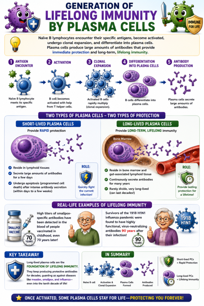
- When naïve B lymphocytes encounter their specific antigen, they become activated.
- The activated B lymphocytes undergo clonal expansion.
- After clonal expansion, they differentiate into:
- Short-lived plasma cells
- Long-lived plasma cells
- Both types of plasma cells produce large amounts of antibodies.
- Short-lived plasma cells provide rapid protection.
- They undergo apoptosis after a few days of intense antibody secretion.
- Long-lived plasma cells remain in the body for a very long time.
- They are mainly located in:
- Bone marrow
- Gut-associated lymphoid tissue (GALT)
- Long-lived plasma cells continue producing antibodies for many years.
- This continuous antibody production provides lifelong immunity against certain infectious diseases.
- Examples include:
- Measles
- Smallpox
- High levels (high titers) of smallpox-specific antibodies have been found in people who were vaccinated 70 years earlier during childhood.
- Older survivors of the 1918 H1N1 influenza pandemic were found to have highly functional virus-neutralizing antibodies even 90 years after infection.
- Therefore, plasma cells producing virus-neutralizing antibodies can survive and function for many decades.
- These plasma cells may continue producing protective antibodies even into the tenth decade of human life.
Key Concept
After activation and clonal expansion, B lymphocytes become either short-lived or long-lived plasma cells. Short-lived plasma cells provide immediate antibody protection, whereas long-lived plasma cells reside mainly in the bone marrow and gut-associated lymphoid tissue, continuously producing antibodies for many years and providing lifelong immunity against diseases such as measles and smallpox.
MADE BY SELF LEARNING CEO AND FOUNDER DR SHEEN
