


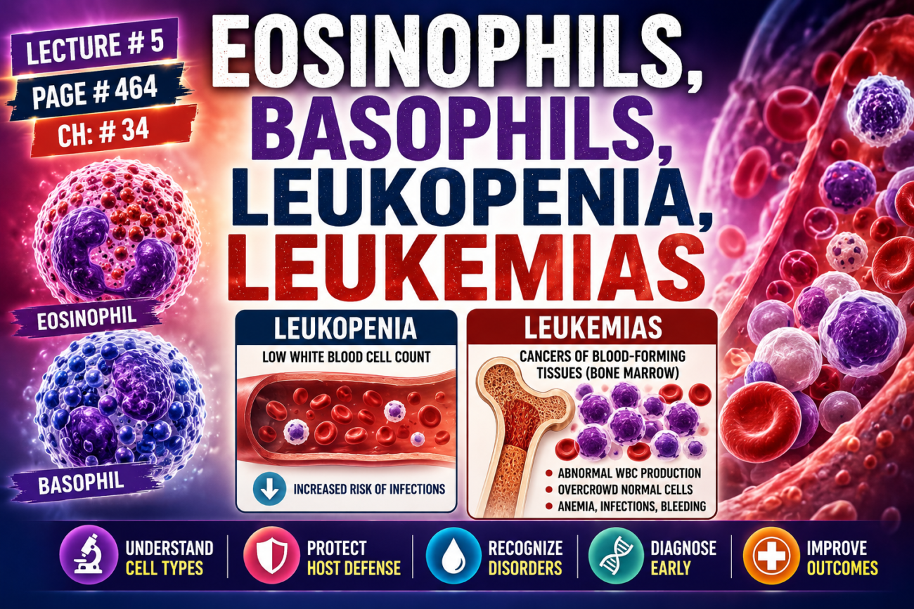
- Eosinophils normally make up about 2% of all blood leukocytes.
- Eosinophils are weak phagocytes.
- They can move toward the site of infection by chemotaxis.
- Compared with neutrophils, eosinophils have only a small role in protecting against common infections.
- Eosinophils are produced in large numbers during parasitic infections.
- They migrate into tissues infected by parasites.
- Most parasites are too large to be engulfed by eosinophils or other phagocytic cells.
- Instead of engulfing them, eosinophils attach to parasites using special surface molecules.
- Eosinophils release substances that kill many parasites.
- Schistosomiasis is one of the most common parasitic infections.
- It occurs in up to one-third of the population in some developing countries of Africa, Asia, and South America.
- About 85%–90% of the world’s cases of schistosomiasis occur in Africa.
- Schistosome worms can invade almost any part of the body.
- Eosinophils attach to the juvenile forms of the parasite.
- They kill many of these parasites.
- Eosinophils kill parasites by releasing hydrolytic enzymes from their granules, which are modified lysosomes.
- They also release highly reactive forms of oxygen that are toxic to parasites.
- They also release a highly larvicidal polypeptide called major basic protein.
- Trichinosis is another parasitic disease that causes eosinophilia.
- Trichinosis occurs when the Trichinella (pork worm) parasite invades the body’s muscles.
- This infection develops after eating undercooked infected pork.
- Eosinophils also collect in tissues where allergic reactions occur.
- They accumulate around the bronchi in the lungs of people with asthma.
- They also collect in the skin during allergic skin reactions.
- This happens because mast cells and basophils participate in allergic reactions.
- Mast cells and basophils release an eosinophil chemotactic factor.
- This factor attracts eosinophils to the allergic tissue.
- Eosinophils help detoxify some inflammatory substances released by mast cells and basophils.
- They also phagocytose and destroy allergen-antibody complexes.
- This helps limit the spread of the local inflammatory response.
- However, excessive and prolonged eosinophil accumulation can worsen breathing problems in people with asthma.
- This occurs because prolonged inflammation causes tissue damage and airway remodeling.
Figure Number
- Figure 34.8: Control of bone marrow production of granulocytes and monocyte-macrophages in response to growth factors released from activated macrophages in inflamed tissue.
Key Concept
- Eosinophils make up about 2% of blood leukocytes and are weak phagocytes.
- Their main role is to defend against parasitic infections and participate in allergic reactions.
- They kill parasites by releasing hydrolytic enzymes, reactive oxygen, and major basic protein.
- Eosinophils also help control allergic inflammation, but excessive accumulation can worsen asthma by causing tissue damage and airway remodeling.
BASOPHILS
- Basophils in the circulating blood are similar to the large mast cells found in tissues.
- Mast cells are located just outside many capillaries in the body.
- Both mast cells and basophils release heparin into the blood.
- Heparin helps prevent blood coagulation (blood clotting).
- Mast cells and basophils also release histamine.
- They also release smaller amounts of bradykinin and serotonin.
- During inflammation, mast cells are the main cells that release these substances.
- Mast cells and basophils play an important role in some types of allergic reactions.
- The antibody immunoglobulin E (IgE) has a strong tendency to attach to mast cells and basophils.
- Later, when the specific antigen reacts with the attached IgE antibody, the mast cell or basophil becomes activated.
- This activation causes the release of large amounts of histamine.
- It also causes the release of bradykinin.
- It also causes the release of serotonin.
- It also causes the release of heparin.
- It also causes the release of slow-reacting substance of anaphylaxis (a mixture of three leukotrienes).
- It also causes the release of several lysosomal enzymes.
- These substances produce local vascular and tissue reactions.
- These reactions are responsible for many, and possibly most, allergic manifestations.
- These allergic reactions are discussed in greater detail in Chapter 35.
Key Concept
- Basophils are similar to tissue mast cells.
- Both cells release heparin, histamine, bradykinin, and serotonin.
- During allergic reactions, IgE activates mast cells and basophils to release inflammatory mediators.
- These mediators produce the vascular and tissue changes responsible for allergic reactions.
LEUKOPENIA
- Leukopenia is a clinical condition in which the bone marrow produces very few white blood cells (WBCs).
- This condition leaves the body poorly protected against bacteria and other invading agents.
- Normally, the human body lives in symbiosis with many bacteria.
- The body’s mucous membranes are constantly exposed to large numbers of bacteria.
- The mouth normally contains spirochetal, pneumococcal, and streptococcal bacteria.
- These bacteria are also present, in smaller numbers, throughout the respiratory tract.
- The distal gastrointestinal tract contains large numbers of colon bacilli.
- Bacteria are also normally present on the surfaces of the eyes, urethra, and vagina.
- When the number of WBCs decreases, these normal bacteria can quickly invade the nearby tissues.
- Within 2 days after the bone marrow stops producing WBCs, ulcers may develop in the mouth and colon.
- Severe respiratory infection may also develop.
- Bacteria from these ulcers rapidly spread into the surrounding tissues and the bloodstream.
- Without treatment, death may occur in less than 1 week after acute total leukopenia begins.
- Exposure to X-rays or gamma rays can cause bone marrow aplasia.
- Drugs and chemicals containing benzene or anthracene nuclei can also cause bone marrow aplasia.
- Some drugs, such as chloramphenicol, may rarely cause leukopenia.
- Thiouracil, used to treat thyrotoxicosis, may also rarely cause leukopenia.
- Some barbiturate hypnotics may also rarely cause leukopenia.
- These causes increase the risk of severe infections.
- After moderate radiation injury, some stem cells, myeloblasts, and hemocytoblasts may survive in the bone marrow.
- These surviving cells can regenerate the bone marrow if enough time is available.
- With proper treatment, including blood transfusions, antibiotics, and other anti-infective drugs, bone marrow recovery is possible.
- Within weeks to months, blood cell counts usually return to normal.
Key Concept
- Leukopenia is a marked decrease in white blood cell production by the bone marrow.
- It greatly increases the risk of severe bacterial infections because normal body bacteria can invade tissues.
- Radiation, certain chemicals, and some drugs can cause leukopenia.
- With appropriate treatment and surviving bone marrow stem cells, normal blood cell production can recover over weeks to months.
LEUKEMIAS
Types of Leukemia
- Uncontrolled production of white blood cells (WBCs) can occur because of a cancerous mutation in a myelogenous or lymphogenous cell.
- This process causes leukemia.
- Leukemia is usually characterized by a greatly increased number of abnormal WBCs in the circulating blood.
- There are two main types of leukemia:
- Lymphocytic leukemia
- Myelogenous leukemia
- Lymphocytic leukemia is caused by cancerous production of lymphoid cells in the bone marrow.
- The abnormal (incompetent) lymphocytes circulate in the blood.
- These lymphocytes accumulate in the lymph nodes and other lymphoid tissues.
- Eventually, they spread to other parts of the body.
- Myelogenous leukemia begins with cancerous production of young myelogenous cells in the bone marrow.
- These abnormal cells spread throughout the body.
- As a result, WBCs are produced in many extramedullary tissues.
- These tissues mainly include the lymph nodes, spleen, and liver.
- In myelogenous leukemia, the cancer cells may sometimes become partially differentiated.
- This may produce:
- Neutrophilic leukemia
- Eosinophilic leukemia
- Basophilic leukemia
- Monocytic leukemia
- More commonly, the leukemia cells are bizarre and undifferentiated.
- These cells do not resemble normal WBCs.
- The more undifferentiated the leukemia cells are, the more acute the leukemia becomes.
- If untreated, acute leukemia may cause death within a few months.
- More differentiated leukemia cells usually produce chronic leukemia.
- Chronic leukemia may develop slowly over 10–20 years.
- Leukemia cells, especially the highly undifferentiated cells, usually cannot provide normal protection against infection.
Key Concept
- Leukemia is a cancer of white blood cells caused by uncontrolled production of abnormal myelogenous or lymphoid cells.
- The two major types are lymphocytic leukemia and myelogenous leukemia.
- Acute leukemia contains highly undifferentiated cells and progresses rapidly, whereas chronic leukemia contains more differentiated cells and progresses slowly.
- Leukemic cells are usually nonfunctional and cannot provide normal protection against infection.
Effects of Leukemia on the Body
- The first effect of leukemia is the metastatic growth of leukemic cells in abnormal areas of the body.
- Leukemic cells from the bone marrow may multiply excessively.
- These cells can invade the surrounding bone.
- This invasion causes bone pain.
- It also increases the tendency for bones to fracture easily.
- Almost all leukemias eventually spread to the spleen.
- They also spread to the lymph nodes.
- They also spread to the liver.
- They also spread to other vascular regions.
- This spread occurs whether the leukemia starts in the bone marrow or the lymph nodes.
- Common effects of leukemia include infection.
- Leukemia also causes severe anemia.
- It also causes a bleeding tendency due to thrombocytopenia (low platelet count).
- These problems occur because nonfunctional leukemic cells replace the normal bone marrow and lymphoid cells.
- Another important effect of leukemia is the excessive use of the body’s metabolic substrates by the growing cancer cells.
- Leukemic tissues produce new cells very rapidly.
- This creates a very high demand for nutrients.
- It also increases the need for specific amino acids.
- It also increases the need for vitamins.
- As a result, the patient’s energy becomes greatly depleted.
- Excessive use of amino acids by leukemic cells causes rapid breakdown of normal body proteins.
- While leukemic tissues continue to grow, the normal body tissues become weak.
- If metabolic starvation continues for a long time, it alone can cause death.
Key Concept
- Leukemia spreads to multiple organs and can invade bones, causing pain and fractures.
- It leads to infection, severe anemia, and thrombocytopenia by replacing normal bone marrow cells.
- Rapid growth of leukemic cells consumes large amounts of nutrients, amino acids, and vitamins, causing weakness, tissue wasting, and eventually death if prolonged.
MADE BY SELF LEARNING CEO AND FOUNDER DR SHEEN

Lectures are explained in very easy way. It becomes so easy to learn and memorize all concepts. Thank you so much
thanks a lot