


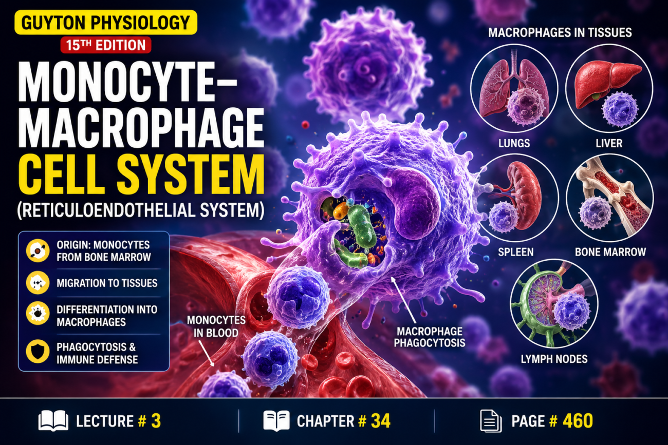
- Macrophages are not only mobile cells that move through the tissues.
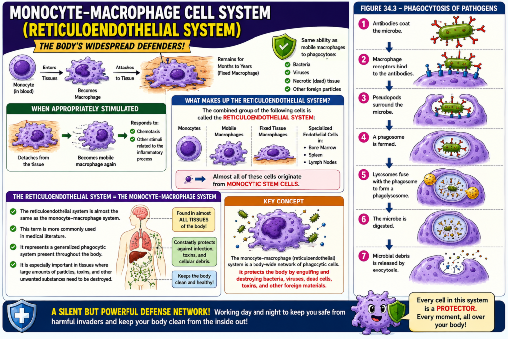
- After entering the tissues and becoming macrophages, many monocytes attach to the tissues.
- These attached macrophages may remain in the tissues for months or even years.
- They perform specific local protective functions.
- Attached macrophages have the same ability as mobile macrophages to phagocytose:
- Bacteria
- Viruses
- Necrotic (dead) tissue
- Other foreign particles
- When appropriately stimulated:
- They detach from the tissues.
- They become mobile macrophages again.
- These mobile macrophages respond to:
- Chemotaxis
- Other stimuli related to the inflammatory process
- Therefore, the body has a widespread monocyte–macrophage system in almost all tissues.
- The combined group of the following cells is called the reticuloendothelial system:
- Monocytes
- Mobile macrophages
- Fixed tissue macrophages
- A few specialized endothelial cells in the:
- Bone marrow
- Spleen
- Lymph nodes
- Almost all of these cells originate from monocytic stem cells.
- Therefore, the reticuloendothelial system is almost the same as the monocyte–macrophage system.
- The term reticuloendothelial system is more commonly used in medical literature.
- It represents a generalized phagocytic system present throughout the body.
- It is especially important in tissues where large amounts of particles, toxins, and other unwanted substances need to be destroyed.
Figure
- Figure 34.3 – Phagocytosis of Pathogens
- Antibodies coat the microbe.
- Macrophage receptors bind to the antibodies.
- Pseudopods surround the microbe.
- A phagosome is formed.
- Lysosomes fuse with the phagosome to form a phagolysosome.
- The microbe is digested.
- Microbial debris is released by exocytosis.
Equations
- No mathematical equation is present in the provided text.
Key Concept
The monocyte–macrophage (reticuloendothelial) system consists of monocytes, mobile macrophages, fixed tissue macrophages, and specialized endothelial cells. Most remain attached to tissues for months to years, perform local phagocytosis, and can become mobile macrophages when stimulated. This system is a body-wide phagocytic defense that removes bacteria, viruses, toxins, dead tissue, and other foreign particles.
Tissue Macrophages in Skin and Subcutaneous Tissues
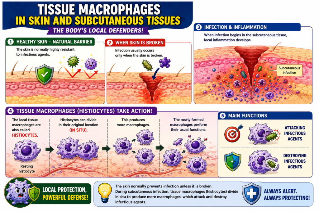
- The skin is normally highly resistant to infectious agents.
- Infection usually occurs only when the skin is broken.
- When infection begins in the subcutaneous tissue, local inflammation develops.
- The local tissue macrophages are also called histiocytes.
- Histiocytes can divide in their original location (in situ).
- This produces more macrophages.
- The newly formed macrophages perform their usual functions.
- These functions include:
- Attacking infectious agents.
- Destroying infectious agents.
Key Concept
The skin normally prevents infection unless it is broken. During subcutaneous infection, tissue macrophages (histiocytes) divide in situ to produce more macrophages, which attack and destroy infectious agents.
Macrophages in Lymph Nodes
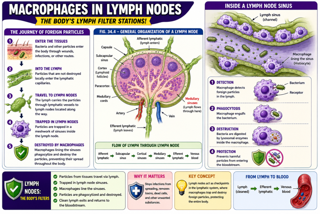
- Particulate matter, such as bacteria, that enters the tissues cannot be absorbed directly into the blood through the capillary membranes.
- If these particles are not destroyed in the tissues, they enter the lymph.
- The particles then travel through the lymph to the lymph nodes located along the lymphatic vessels.
- Inside the lymph nodes, the foreign particles become trapped in a meshwork of sinuses.
- These sinuses are lined with tissue macrophages.
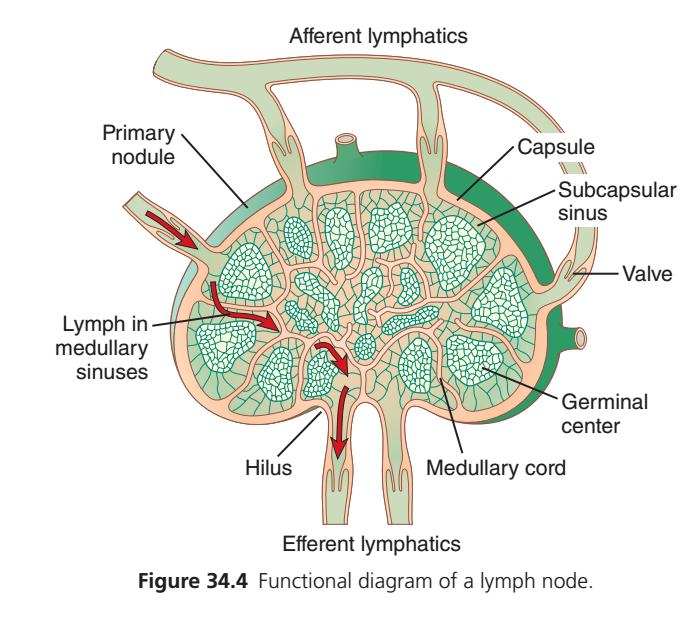
- Figure 34.4 shows the general structure of a lymph node.
- Lymph enters the lymph node through the capsule by afferent lymphatic vessels.
- It then flows through the medullary sinuses of the lymph node.
- Finally, the lymph leaves the hilus through the efferent lymphatic vessels.
- The efferent lymphatic vessels eventually empty into the venous blood.
- Large numbers of macrophages line the lymph sinuses.
- If foreign particles enter the sinuses with the lymph, the macrophages phagocytose them.
- This prevents the spread (general dissemination) of foreign particles throughout the body.
Figure
- Figure 34.4 – General Organization of a Lymph Node
- Lymph enters through afferent lymphatics.
- Lymph passes through the lymph node capsule.
- Lymph flows through the medullary sinuses.
- Lymph exits through the hilus.
- Lymph leaves via efferent lymphatics.
- Macrophages line the lymph sinuses and phagocytose foreign particles.
Key Concept
Lymph nodes act as filters for the lymph. Macrophages lining the lymph sinuses trap and phagocytose bacteria and other foreign particles, preventing their spread into the bloodstream and throughout the body.
Structure and Function of a Lymph Node (Guyton Figure 34.4)
One Main Idea
A lymph node is a small biological filter that cleans the lymph before it returns to the bloodstream.
It traps:
- 🦠 Bacteria
- 🦠 Viruses
- 🧫 Foreign particles
- 💀 Dead cells
- 🎗️ Cancer cells
At the same time, it activates the immune system to fight infection.
Easy Definition:
Lymph Node = Body’s Security Checkpoint + Water Filter
Easy Flow of Lymph Through a Lymph Node
Tissues
    ↓
Afferent Lymphatic Vessels
    ↓
Subcapsular Sinus
    ↓
Cortex (Nodules)
    ↓
Medullary Sinuses
    ↓
Hilum
    ↓
Efferent Lymphatic Vessel
    ↓
Blood Circulation
Step 1. Afferent Lymphatic Vessels (Entry)
What happens?
Lymph coming from body tissues enters the lymph node through several afferent lymphatic vessels.
The lymph carries:
- Bacteria
- Viruses
- Foreign particles
- Tissue fluid
Easy Memory
Afferent = Arrives
➡️ Lymph arrives at the lymph node.
Step 2. Capsule
The entire lymph node is covered by a tough outer layer called the capsule.
Function
- Protects the lymph node
- Maintains its shape
Easy Analogy
Think of the capsule as the outer wall of a security building.
Step 3. Subcapsular Sinus
Just beneath the capsule is a large space called the subcapsular sinus.
Function
This is the first area where incoming lymph flows.
Here:
- Lymph slows down.
- Macrophages begin inspecting it.
Easy Concept
This is the first security checkpoint.
Step 4. Cortex (Primary Nodules)
The outer part of the lymph node contains many primary nodules.
These nodules contain mostly:
🟢 B lymphocytes
Function
They continuously monitor lymph for invading microorganisms.
Step 5. Germinal Center
When bacteria or viruses enter:
The primary nodule becomes active.
Its center enlarges to form a:
Germinal Center
Function
Here, B cells rapidly divide and mature into:
- Plasma cells
- Memory B cells
Plasma cells produce antibodies.
Easy Memory
Germinal Center = B-cell factory
Step 6. Medullary Cord
The inner region of the lymph node contains the medullary cords.
They contain:
- Plasma cells
- Macrophages
- B lymphocytes
- T lymphocytes
Function
These cells continue destroying pathogens and producing antibodies.
Easy Concept
The medullary cords are the immune army headquarters.
Step 7. Medullary Sinuses
Between the medullary cords are open channels called the medullary sinuses.
Function
Lymph slowly flows through these channels.
While passing:
- Macrophages trap bacteria.
- Foreign particles are removed.
Easy Concept
These sinuses act like the final filtration channels.
Step 8. Hilum
The hilum is the gateway of the lymph node.
Here:
- Clean lymph leaves.
- Blood vessels enter and leave the node.
Easy Memory
Hilum = Exit Gate
Step 9. Efferent Lymphatic Vessel (Exit)
Finally, filtered lymph leaves through the efferent lymphatic vessel.
Easy Memory
Efferent = Exit
Whole Process in One Flow
Body tissues
        ↓
Afferent lymphatic vessels
        ↓
Subcapsular sinus
        ↓
Primary nodules
        ↓
Germinal centers activated
        ↓
Medullary cords
        ↓
Medullary sinuses
        ↓
Hilum
        ↓
Efferent lymphatic vessel
        ↓
Clean lymph returns to blood
Easy Analogy
Imagine an airport security system.
- 🚶 Lymph = Passengers
- 🧳 Bacteria & viruses = Dangerous items
- 👮 Macrophages = Security officers
- 🛡️ Lymph node = Security checkpoint
- 🏭 Germinal center = Factory producing antibodies
- 🚪 Efferent vessel = Exit gate
Every passenger (lymph) is checked before being allowed to leave.
Functions of a Lymph Node
✅ Filters lymph
✅ Traps bacteria and viruses
✅ Removes dead cells and foreign particles
✅ Houses macrophages for phagocytosis
✅ Activates B and T lymphocytes
✅ Produces antibodies through plasma cells
Important Structures and Their Functions
| Structure | Function |
|---|---|
| Afferent lymphatic vessel | Brings lymph into the lymph node. |
| Capsule | Protective outer covering of the lymph node. |
| Subcapsular sinus | First chamber where incoming lymph is filtered. |
| Primary nodule | Contains resting B lymphocytes. |
| Germinal center | Activated B-cell area where B cells proliferate and differentiate into plasma and memory cells. |
| Medullary cord | Contains plasma cells, macrophages, B cells, and T cells involved in immune responses. |
| Medullary sinuses | Channels through which lymph flows and is filtered by macrophages. |
| Hilum | Exit region for lymph and entry/exit point for blood vessels. |
| Efferent lymphatic vessel | Carries filtered lymph away from the lymph node. |
Memory Tricks
Afferent = Arrives
Lymph enters the node.
Efferent = Exit
Lymph leaves the node.
Germinal Center = Antibody Factory
B cells multiply and become plasma cells.
Hilum = Highway Exit
The final exit for filtered lymph.
High-Yield Exam Points (Guyton)
- Lymph nodes act as biological filters, trapping microorganisms, foreign particles, and cellular debris before lymph returns to the bloodstream.
- Afferent lymphatic vessels bring lymph into the node, while the efferent lymphatic vessel carries filtered lymph away.
- The subcapsular sinus is the first region through which incoming lymph flows.
- Primary nodules contain resting B lymphocytes.
- During infection, germinal centers develop, where B cells proliferate and differentiate into plasma cells and memory B cells.
- Medullary cords contain plasma cells, macrophages, B lymphocytes, and T lymphocytes.
- Medullary sinuses provide additional filtration as macrophages remove pathogens and debris.
- The hilum is the exit point for filtered lymph and the entry/exit site for blood vessels.
Alveolar Macrophages in Lungs
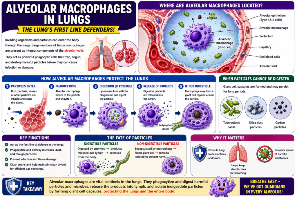
- Another route for infectious organisms to enter the body is through the lungs.
- Large numbers of tissue macrophages are present in the alveolar walls.
- These are called alveolar macrophages.
- Alveolar macrophages phagocytose particles that become trapped in the alveoli.
- If the particles are digestible:
- The macrophages digest them.
- The digestive products are released into the lymph.
- If the particles are not digestible:
- The macrophages form a giant cell capsule around the particle.
- The capsule remains around the particle until it is slowly dissolved, if it can be dissolved.
- Giant cell capsules are commonly formed around:
- Tuberculosis bacilli
- Silica dust particles
- Carbon particles
Key Concept
Alveolar macrophages are located in the alveolar walls and protect the lungs by phagocytosing inhaled particles. Digestible particles are broken down and released into the lymph, while indigestible particles are enclosed within a giant cell capsule, such as those formed around tuberculosis bacilli, silica dust, and carbon particles.
Macrophages (Kupffer Cells) in Liver Sinusoids
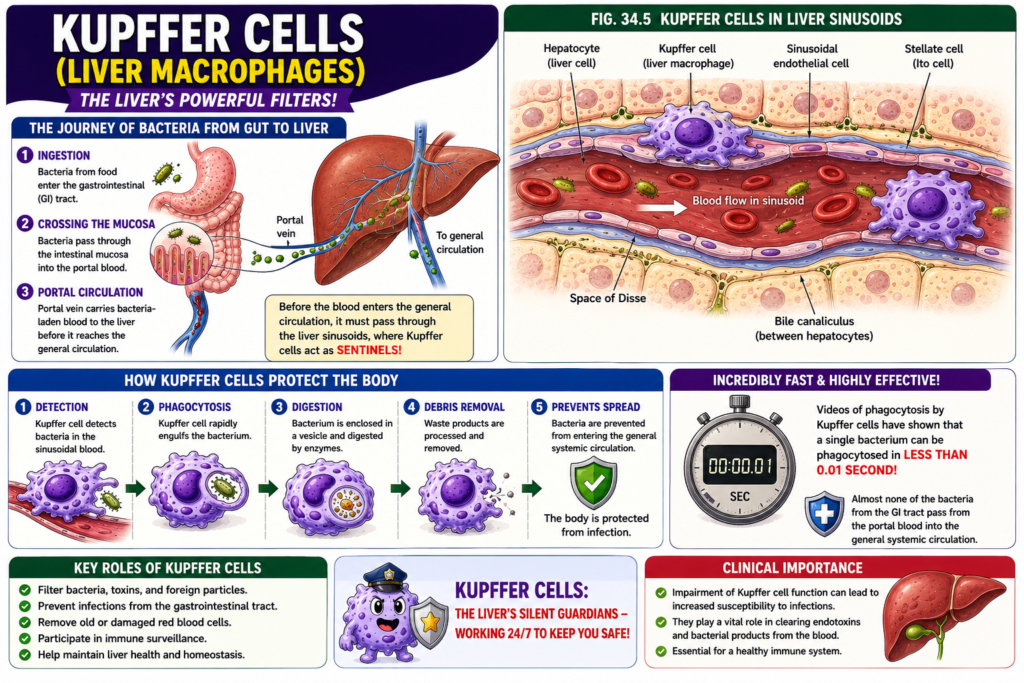
- Another route by which bacteria enter the body is through the gastrointestinal (GI) tract.
- Large numbers of bacteria from ingested food continuously pass through the gastrointestinal mucosa into the portal blood.
- Before the portal blood enters the general circulation, it passes through the liver sinusoids.
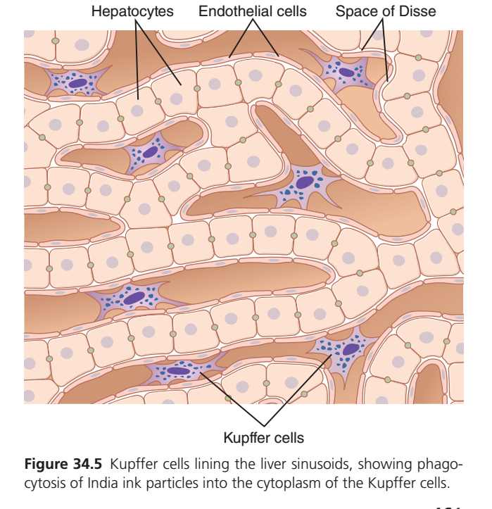
- The liver sinusoids are lined with tissue macrophages called Kupffer cells.
- Kupffer cells are shown in Figure 34.5.
- Kupffer cells form a highly effective particulate filtration system.
- They remove bacteria from the portal blood.
- As a result, almost no bacteria from the gastrointestinal tract enter the general systemic circulation.
- Videos of Kupffer cells have shown that they can phagocytose a single bacterium in less than 0.01 second.
Figure
- Figure 34.5 – Kupffer cells lining the liver sinusoids, filtering bacteria from the portal blood before it enters the systemic circulation.
Key Concept
Kupffer cells are macrophages located in the liver sinusoids. They act as an efficient filtration system, removing bacteria from the portal blood before it reaches the systemic circulation, and can phagocytose a bacterium in less than 0.01 second.
Macrophages of Spleen and Bone Marrow
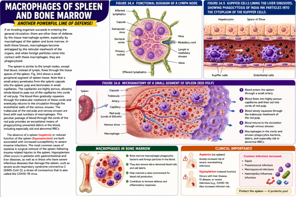
- If an infectious organism enters the general circulation, the tissue macrophage system provides another line of defense.
- This defense is mainly provided by macrophages in the spleen and bone marrow.
- In both the spleen and bone marrow, macrophages are trapped within the reticular meshwork.
- When foreign particles come into contact with these macrophages, they are phagocytosed.
- The spleen is similar to the lymph nodes.
- The difference is that blood, instead of lymph, flows through the tissue spaces of the spleen.
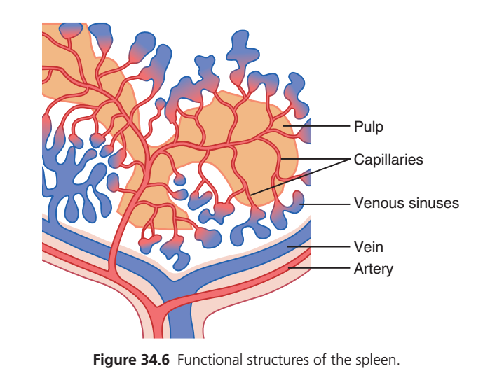
- Figure 34.6 shows a small peripheral segment of spleen tissue.
- A small artery enters the spleen through the splenic capsule.
- The artery ends in small capillaries.
- These capillaries are highly porous.
- Whole blood passes out of the capillaries into the cords of the red pulp.
- The blood then slowly passes through the trabecular meshwork of the red pulp.
- Finally, the blood returns to the circulation through the endothelial walls of the venous sinuses.
- The trabeculae of the red pulp and the venous sinuses are lined with large numbers of macrophages.
- This special blood flow through the red pulp allows efficient phagocytosis of unwanted materials in the blood.
- The macrophages especially remove:
- Old red blood cells (RBCs)
- Abnormal red blood cells (RBCs)
- Absence of the spleen (asplenia) increases the risk of severe invasive infections.
- Reduced spleen function (hyposplenism) also increases susceptibility to severe invasive infections.
- The most common cause of asplenia is surgical removal of the spleen after trauma-related injury.
- Hyposplenism commonly occurs in patients with:
- Gastrointestinal diseases
- Liver diseases
- Severe infectious diseases that damage the spleen
- One example is infection with severe acute respiratory syndrome coronavirus 2 (SARS-CoV-2), also called the COVID-19 virus.
Figure
- Figure 34.4 – Functional Diagram of a Lymph Node
- Afferent lymphatics
- Capsule
- Subcapsular sinus
- Germinal center
- Primary nodule
- Medullary cord
- Lymph in medullary sinuses
- Hilus
- Efferent lymphatics
- Valve
- Figure 34.5 – Kupffer Cells in Liver Sinusoids
- Kupffer cells
- Hepatocytes
- Endothelial cells
- Space of Disse
- Phagocytosis of India ink particles by Kupffer cells
- Figure 34.6 – Peripheral Segment of Spleen Tissue
- Splenic capsule
- Small artery
- Porous capillaries
- Red pulp cords
- Trabecular meshwork
- Venous sinuses
- Macrophages lining the red pulp and venous sinuses
Key Concept
Macrophages in the spleen and bone marrow provide an important blood-filtering defense by phagocytosing foreign particles, especially old and abnormal RBCs. The spleen filters blood (not lymph) through the red pulp, where numerous macrophages remove unwanted materials. Asplenia and hyposplenism increase the risk of severe invasive infections.
MADE BY SELF LEARNING CEO AND FOUNDER DR SHEEN
