


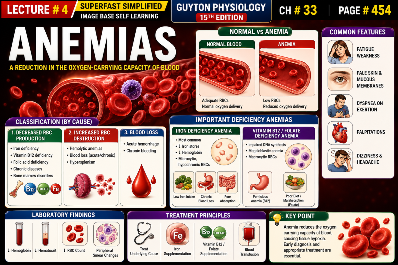
- Anemia means a deficiency of hemoglobin in the blood.
- Anemia can occur because:
- There are too few RBCs.
- The RBCs contain too little hemoglobin.
- Different types of anemia have different physiological causes.
- Blood Loss Anemia
- After a rapid hemorrhage, the body replaces the fluid portion of the plasma within 1–3 days.
- This causes the concentration of RBCs in the blood to become low.
- If no second hemorrhage occurs:
- The RBC concentration usually returns to normal within 3–6 weeks.
- During chronic blood loss:
- The body often cannot absorb enough iron from the intestines.
- As a result, hemoglobin cannot be produced as rapidly as it is lost.
- Consequently:
- RBCs become much smaller than normal.
- RBCs contain too little hemoglobin.
- This condition produces microcytic hypochromic anemia.
- Figure 33.3 shows microcytic hypochromic anemia.
KEY CONCEPT
- Anemia is a deficiency of hemoglobin in the blood.
- It results from:
- Too few RBCs, or
- Too little hemoglobin in RBCs.
- After acute hemorrhage, plasma volume is restored in 1–3 days, while RBC concentration returns to normal in 3–6 weeks if no further bleeding occurs.
- Chronic blood loss causes iron deficiency, reducing hemoglobin production.
- This leads to microcytic hypochromic anemia, in which RBCs are small and contain less hemoglobin.
- Figure 33.3 illustrates microcytic hypochromic anemia.
ANEMIAS
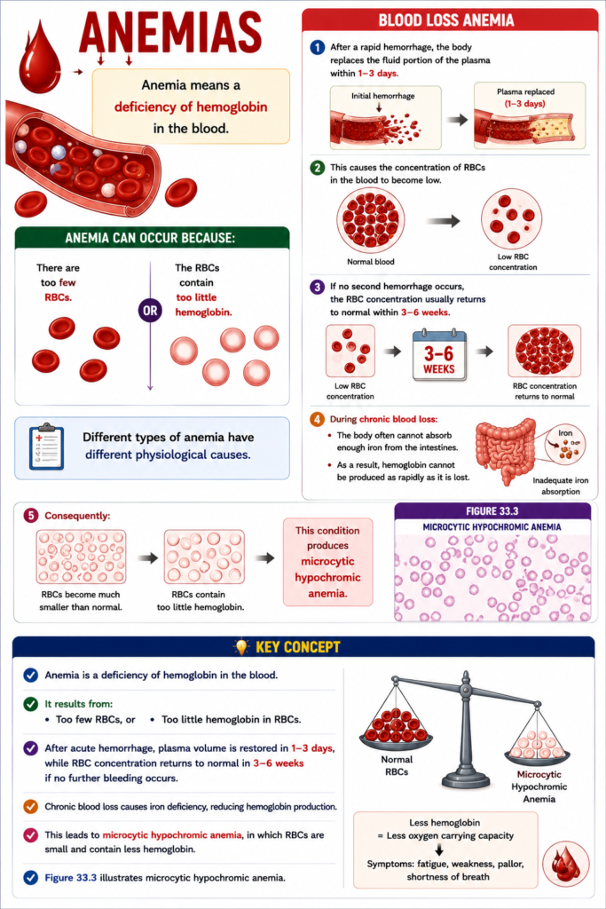
- Aplastic anemia is caused by bone marrow dysfunction.
- Bone marrow aplasia means lack of functioning bone marrow.
- Exposure to high-dose radiation can damage bone marrow stem cells.
- Chemotherapy for cancer treatment can also damage bone marrow stem cells.
- A few weeks after stem cell damage, anemia develops.
- High doses of certain toxic chemicals can also damage the bone marrow.
- Examples include:
- Insecticides
- Benzene in gasoline
- These toxic chemicals can produce the same effect as radiation and chemotherapy.
- In autoimmune disorders, the immune system attacks healthy cells.
- In lupus erythematosus, the immune system may attack bone marrow stem cells.
- This can lead to aplastic anemia.
- In about half of all aplastic anemia cases, the cause is unknown.
- This condition is called idiopathic aplastic anemia.
- People with severe aplastic anemia usually die if they are not treated.
- Blood transfusions can temporarily increase the number of RBCs.
- Bone marrow transplantation is another treatment option.
KEY CONCEPT
- Aplastic anemia results from bone marrow dysfunction.
- Bone marrow aplasia means non-functioning bone marrow.
- Causes include:
- High-dose radiation
- Chemotherapy
- Toxic chemicals (insecticides, benzene)
- Autoimmune disorders such as lupus erythematosus
- About 50% of cases are idiopathic (cause unknown).
- Severe aplastic anemia requires treatment with blood transfusions or bone marrow transplantation.
ANEMIAS
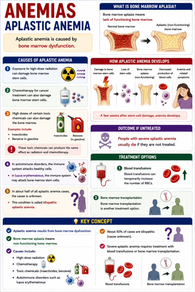
- Megaloblastic anemia is characterized by abnormally large, poorly developed RBCs.
- Deficiency of any of the following can slow the reproduction of erythroblasts in the bone marrow:
- Vitamin B12
- Folic acid
- Intrinsic factor
- As a result, RBCs grow too large.
- These abnormally large RBCs have irregular (odd) shapes.
- These cells are called megaloblasts.
- Atrophy of the stomach mucosa, as seen in pernicious anemia, can cause megaloblastic anemia.
- Total gastrectomy (complete surgical removal of the stomach) can also lead to megaloblastic anemia.
- Megaloblastic anemia also commonly develops in patients with intestinal sprue.
- In intestinal sprue, absorption of the following is poor:
- Folic acid
- Vitamin B12
- Other vitamin B compounds
- Because erythroblasts cannot proliferate rapidly enough, they cannot produce a normal number of RBCs.
- The RBCs that are produced are:
- Mostly oversized
- Bizarre in shape
- Have fragile membranes
- These abnormal RBCs rupture easily.
- As a result, the person develops a severe shortage of adequate RBCs.
KEY CONCEPT
- Megaloblastic anemia is caused by abnormally large, poorly developed RBCs.
- Deficiency of vitamin B12, folic acid, or intrinsic factor slows erythroblast reproduction.
- Slow cell division produces large abnormal RBCs (megaloblasts).
- Causes include:
- Pernicious anemia (atrophy of the stomach mucosa)
- Total gastrectomy
- Intestinal sprue with poor absorption of folic acid, vitamin B12, and other vitamin B compounds
- The resulting RBCs are oversized, irregularly shaped, and have fragile membranes.
- These fragile cells rupture easily, leading to anemia.
ANEMIAS
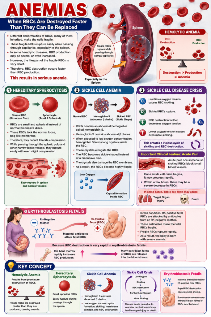
- Hemolytic anemia occurs when RBCs are destroyed faster than they can be replaced.
- Different abnormalities of RBCs, many of them inherited, make the cells fragile.
- These fragile RBCs rupture easily while passing through capillaries, especially in the spleen.
- In some hemolytic diseases, RBC production may be normal or even increased.
- However, the lifespan of the fragile RBCs is very short.
- Therefore, RBC destruction occurs faster than RBC production.
- This results in serious anemia.
- Hereditary spherocytosis
- RBCs are small and spherical instead of normal biconcave discs.
- These RBCs lack the normal loose, bag-like membrane.
- Therefore, they cannot tolerate compression.
- While passing through the splenic pulp and other narrow blood vessels, they rupture easily with even slight compression.
- Sickle cell anemia
- RBCs contain an abnormal hemoglobin called hemoglobin S.
- Hemoglobin S contains abnormal beta (β) chains.
- When exposed to low oxygen concentration, hemoglobin S forms long crystals inside the RBC.
- These crystals elongate the RBC.
- The RBC becomes sickle-shaped instead of a biconcave disc.
- The crystals also damage the RBC membrane.
- As a result, the RBCs become highly fragile.
- This leads to serious anemia.
- Sickle cell disease crisis
- Low tissue oxygen tension causes RBC sickling.
- Sickled RBCs rupture.
- RBC destruction further decreases oxygen tension.
- Lower oxygen tension causes even more sickling.
- This creates a vicious cycle of sickling and RBC destruction.
- An important clinical feature of sickle cell crisis is acute pain.
- Acute pain occurs because sickled RBCs block small blood vessels.
- Once sickle cell crisis begins, it may progress rapidly.
- Within a few hours, there may be a severe decrease in RBCs.
- In some cases, sickle cell crisis may cause:
- Target organ injury
- Death
- Erythroblastosis fetalis
- In this condition, Rh-positive fetal RBCs are attacked by antibodies from an Rh-negative mother.
- These antibodies make the fetal RBCs fragile.
- Fragile RBCs rupture rapidly.
- As a result, the baby is born with severe anemia.
- Because RBC destruction is very rapid in erythroblastosis fetalis:
- The bone marrow rapidly increases RBC production.
- Many early blast forms of RBCs are released into the bloodstream.
KEY CONCEPT
- Hemolytic anemia results from premature destruction of RBCs.
- Fragile RBCs are destroyed faster than they are produced, causing anemia.
- Hereditary spherocytosis:
- Small, spherical RBCs
- Easily rupture during passage through the spleen.
- Sickle cell anemia:
- Hemoglobin S contains abnormal β chains.
- Low oxygen causes crystal formation, sickling, membrane damage, and RBC destruction.
- Sickle cell crisis:
- Low oxygen → Sickling → RBC destruction → Further low oxygen → More sickling.
- Causes acute pain due to vascular occlusion and may lead to organ injury or death.
- Erythroblastosis fetalis:
- Maternal antibodies destroy Rh-positive fetal RBCs.
- Rapid RBC destruction causes severe anemia.
- Bone marrow releases many immature blast forms of RBCs into the blood.
Effects of Anemia on Circulatory System Function
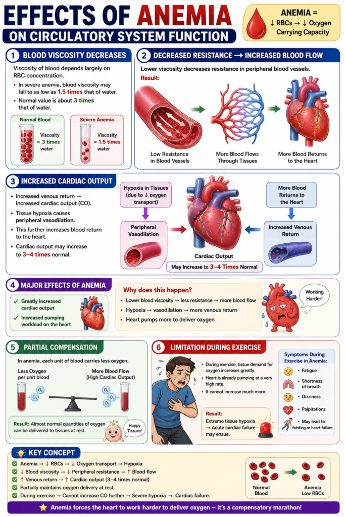
- Blood viscosity depends mainly on the concentration of RBCs.
- In people with severe anemia, blood viscosity may decrease to about 1.5 times the viscosity of water.
- Normally, blood viscosity is about 3 times the viscosity of water.
- Lower blood viscosity decreases the resistance to blood flow in the peripheral blood vessels.
- As a result, much larger amounts of blood flow through the tissues.
- More blood also returns to the heart.
- This greatly increases cardiac output.
- Reduced oxygen transport in anemia causes hypoxia.
- Hypoxia causes the peripheral blood vessels to dilate.
- Vasodilation allows even more blood to return to the heart.
- This further increases cardiac output.
- Cardiac output may increase to 3–4 times the normal value.
- Therefore, one of the major effects of anemia is a marked increase in cardiac output.
- The increased cardiac output also increases the pumping workload of the heart.
- The increased cardiac output partly compensates for the reduced oxygen-carrying capacity of the blood.
- Although each unit of blood carries less oxygen, the higher blood flow helps deliver almost normal amounts of oxygen to the tissues.
- During exercise, tissue oxygen demand increases greatly.
- In a person with anemia, the heart is already pumping at a high rate.
- Therefore, the heart cannot increase cardiac output much further during exercise.
- As a result, severe tissue hypoxia develops during exercise.
- Severe tissue hypoxia may lead to acute cardiac failure.
KEY CONCEPT
- Blood viscosity depends mainly on the RBC concentration.
- In severe anemia:
- Blood viscosity falls from about 3 to 1.5 times the viscosity of water.
- Peripheral vascular resistance decreases.
- Blood flow to tissues and venous return increase.
- Hypoxia causes peripheral vasodilation, further increasing venous return and cardiac output.
- Cardiac output may rise to 3–4 times normal.
- Increased cardiac output partially compensates for reduced oxygen-carrying capacity.
- During exercise, the heart cannot increase output much further, leading to severe tissue hypoxia and possible acute cardiac failure.
POLYCYTHEMIA
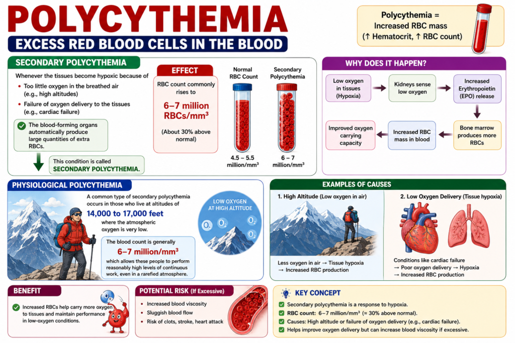
- Secondary polycythemia occurs when the tissues become hypoxic.
- Tissue hypoxia may occur because:
- Too little oxygen is present in the inspired air, such as at high altitudes.
- Oxygen delivery to the tissues is reduced, such as in cardiac failure.
- In response to hypoxia, the blood-forming organs automatically produce large numbers of additional RBCs.
- This increase in RBC production is called secondary polycythemia.
- In secondary polycythemia, the RBC count commonly increases to 6–7 million/mm³.
- This is about 30% above the normal RBC count.
- A common type of secondary polycythemia is physiological polycythemia.
- Physiological polycythemia occurs in people living at 14,000–17,000 feet above sea level.
- At these high altitudes, the atmospheric oxygen level is very low.
- The RBC count in these people is generally 6–7 million/mm³.
- This increased RBC count allows them to perform reasonably high levels of continuous work.
- They are able to do this even in a rarefied (low-oxygen) atmosphere.
KEY CONCEPT
- Secondary polycythemia develops due to tissue hypoxia.
- Causes of hypoxia include:
- High altitude
- Cardiac failure
- Hypoxia stimulates the blood-forming organs to produce more RBCs.
- The RBC count commonly rises to 6–7 million/mm³, about 30% above normal.
- Physiological polycythemia occurs in people living at 14,000–17,000 feet, where atmospheric oxygen is low.
- The increased RBC count helps maintain work capacity in a low-oxygen (rarefied) environment.
POLYCYTHEMIA
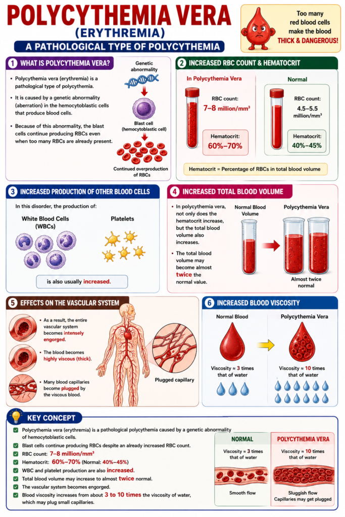
- Polycythemia vera (erythremia) is a pathological type of polycythemia.
- It is caused by a genetic abnormality (aberration) in the hemocytoblastic cells that produce blood cells.
- Because of this abnormality, the blast cells continue producing RBCs even when too many RBCs are already present.
- In polycythemia vera, the RBC count may increase to 7–8 million/mm³.
- The hematocrit may increase to 60%–70%.
- The normal hematocrit is 40%–45%.
- In this disorder, the production of:
- White blood cells (WBCs)
- Platelets
- In polycythemia vera, not only does the hematocrit increase, but the total blood volume also increases.
- The total blood volume may become almost twice the normal value.
- As a result, the entire vascular system becomes intensely engorged.
- The blood becomes highly viscous (thick).
- Many blood capillaries become plugged by the viscous blood.
- Normally, blood viscosity is about 3 times the viscosity of water.
- In polycythemia vera, blood viscosity may increase to about 10 times the viscosity of water.
KEY CONCEPT
- Polycythemia vera (erythremia) is a pathological polycythemia caused by a genetic abnormality of hemocytoblastic cells.
- Blast cells continue producing RBCs despite an already increased RBC count.
- RBC count: 7–8 million/mm³
- Hematocrit: 60%–70% (Normal: 40%–45%)
- WBC and platelet production are also increased.
- Total blood volume may increase to almost twice normal.
- The vascular system becomes engorged.
- Blood viscosity increases from about 3 to 10 times the viscosity of water, which may plug small capillaries.
Effect of Polycythemia on Function of the Circulatory System
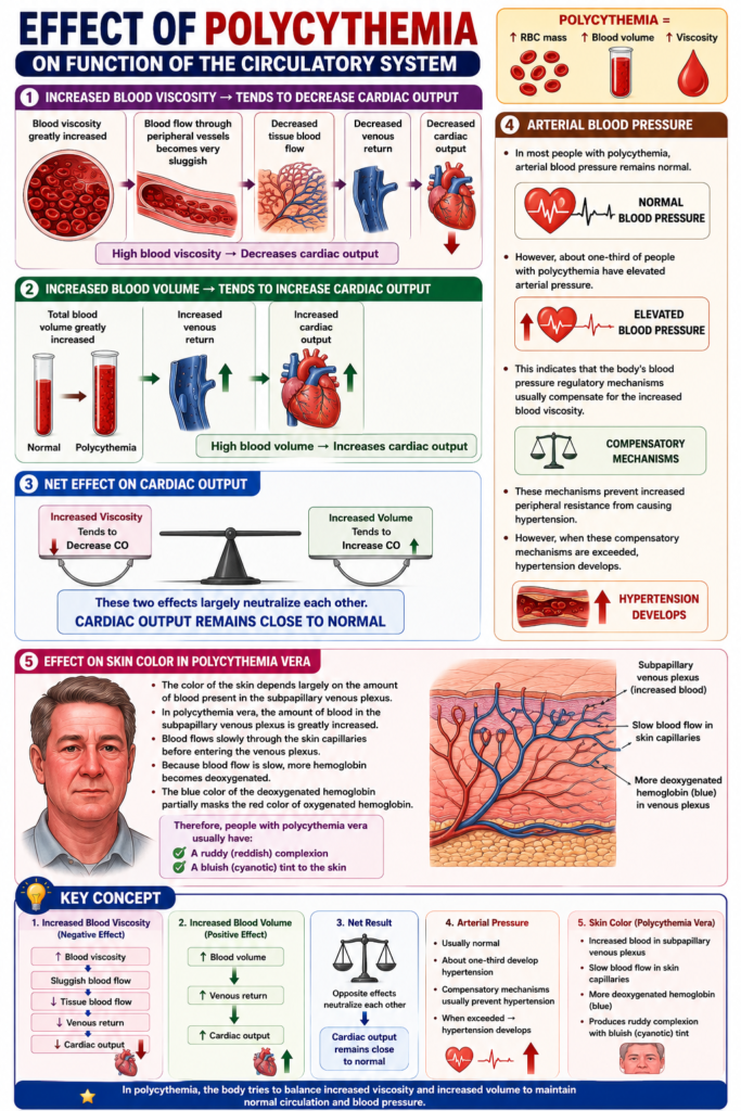
- In polycythemia, blood viscosity is greatly increased.
- Because the blood is more viscous, blood flow through the peripheral blood vessels becomes very sluggish.
- Increased blood viscosity decreases tissue blood flow.
- Reduced tissue blood flow decreases venous return.
- Reduced venous return decreases cardiac output.
- However, in polycythemia, the total blood volume is greatly increased.
- The increased blood volume increases venous return.
- The increased blood volume also increases cardiac output.
- Therefore, in polycythemia:
- Increased blood viscosity tends to decrease cardiac output.
- Increased blood volume tends to increase cardiac output.
- These two effects largely neutralize each other.
- As a result, cardiac output remains close to normal.
- In most people with polycythemia, arterial blood pressure remains normal.
- However, about one-third of people with polycythemia have elevated arterial pressure.
- This indicates that the body’s blood pressure regulatory mechanisms usually compensate for the increased blood viscosity.
- These mechanisms prevent increased peripheral resistance from causing hypertension.
- However, when these compensatory mechanisms are exceeded, hypertension develops.
- The color of the skin depends largely on the amount of blood present in the subpapillary venous plexus.
- In polycythemia vera, the amount of blood in the subpapillary venous plexus is greatly increased.
- Blood flows slowly through the skin capillaries before entering the venous plexus.
- Because blood flow is slow, more hemoglobin becomes deoxygenated.
- The blue color of the deoxygenated hemoglobin partially masks the red color of oxygenated hemoglobin.
- Therefore, people with polycythemia vera usually have:
- A ruddy (reddish) complexion
- A bluish (cyanotic) tint to the skin
KEY CONCEPT
- Increased blood viscosity in polycythemia causes sluggish blood flow.
- High blood viscosity:
- Decreases tissue blood flow
- Decreases venous return
- Decreases cardiac output
- Increased blood volume:
- Increases venous return
- Increases cardiac output
- These opposite effects keep cardiac output near normal.
- Arterial pressure is usually normal, but about one-third of patients develop hypertension when compensatory mechanisms fail.
- In polycythemia vera, increased blood in the subpapillary venous plexus and increased deoxygenated hemoglobin produce a ruddy complexion with a bluish (cyanotic) tint.
MADE BY SELF LEARNING CEO AND FOUNDER DR SHEEN
