


- The second line of defense against acid–base disturbances is the lungs.
- The lungs regulate the CO₂ concentration in the extracellular fluid.
- Increased ventilation removes more CO₂ from the extracellular fluid.
- Removal of CO₂ decreases H⁺ concentration by mass action.
- Decreased H⁺ concentration increases pH.
- Decreased ventilation causes CO₂ to accumulate.
- Increased CO₂ increases H⁺ concentration.
- Increased H⁺ concentration decreases pH.
Easy Concept
↑ Ventilation
↓
↓ CO₂
↓
↓ H₂CO₃
↓
↓ H⁺
↓
↑ pH (Alkalosis)
↓ Ventilation
↓
↑ CO₂
↓
↑ H₂CO₃
↓
↑ H⁺
↓
↓ pH (Acidosis)
Pulmonary Expiration of CO₂ Balances Metabolic Formation of CO₂
- CO₂ is continuously produced inside body cells.
- CO₂ is formed during intracellular metabolism.
- CO₂ diffuses from cells into the interstitial fluid.
- CO₂ then diffuses into the blood.
- Blood transports CO₂ to the lungs.
- CO₂ diffuses from blood into the alveoli.
- Pulmonary ventilation removes CO₂ into the atmosphere.
- Normally, the extracellular fluid contains about 1.2 mmol/L of dissolved CO₂.
- This corresponds to a PCO₂ of 40 mm Hg.
Mathematical Relationship
1.2 mmol/L dissolved CO₂⟺PCO2=40 mm Hg
Easy Concept
Cells
↓
Produce CO₂
↓
Blood carries CO₂
↓
Lungs remove CO₂
↓
Body maintains normal PCO₂ = 40 mm Hg
- If metabolic CO₂ production increases, extracellular PCO₂ increases.
- If metabolic CO₂ production decreases, extracellular PCO₂ decreases.
- Increasing pulmonary ventilation removes more CO₂ from the lungs.
- Increased ventilation decreases extracellular PCO₂.
- Therefore, extracellular PCO₂ depends on:
- Pulmonary ventilation.
- Tissue CO₂ production.
Easy Concept
More Tissue Metabolism
↓
More CO₂
↓
Higher PCO₂
More Ventilation
↓
More CO₂ Removed
↓
Lower PCO₂
Increasing Alveolar Ventilation Decreases Extracellular Fluid H⁺ Concentration and Raises pH
Figure Mentioned: Fig. 31.2
- If metabolic CO₂ production remains constant, alveolar ventilation is the only factor that changes extracellular PCO₂.
- Higher alveolar ventilation produces a lower PCO₂.
- Increased CO₂ increases H₂CO₃ concentration.
- Increased H₂CO₃ increases H⁺ concentration.
- Increased H⁺ lowers extracellular fluid pH.
Easy Concept
↑ CO₂
↓
↑ H₂CO₃
↓
↑ H⁺
↓
↓ pH
- Fig. 31.2 shows the changes in blood pH produced by changing alveolar ventilation.
- Increasing alveolar ventilation to 2 times normal raises extracellular fluid pH by about 0.23.
Mathematical Solution
Normal pH7.40
Increase in pH+0.23
Final pH7.40+0.23=7.63
Final Answer
pH=7.63
- Therefore, if normal pH is 7.40, doubling alveolar ventilation raises the pH to about 7.63.
Easy Concept
Ventilation ×2
↓
CO₂ decreases
↓
H⁺ decreases
↓
pH increases
↓
7.40 → 7.63
- Decreasing alveolar ventilation to one-fourth normal lowers pH by about 0.45.
Mathematical Solution
Normal pH7.40
Decrease in pH−0.45
Final pH7.40−0.45=6.95
Final Answer
pH=6.95
- Therefore, reducing alveolar ventilation to one-fourth normal decreases the pH from 7.40 to 6.95.
Easy Concept
Ventilation = ¼ Normal
↓
CO₂ increases
↓
H⁺ increases
↓
pH decreases
↓
7.40 → 6.95
- Alveolar ventilation can change over a very wide range.
- It may decrease to 0.
- It may increase up to 15 times normal.
- Therefore, the respiratory system can produce large changes in body fluid pH.
Easy Concept
Ventilation Range0⟶15×Normal
↓
Large changes in CO₂
↓
Large changes in H⁺
↓
Large changes in pH
KEY CONCEPT
- Respiratory system is the second line of defense against acid–base disorders.
- ↑ Ventilation → ↓ CO₂ → ↓ H₂CO₃ → ↓ H⁺ → ↑ pH (Respiratory Alkalosis).
- ↓ Ventilation → ↑ CO₂ → ↑ H₂CO₃ → ↑ H⁺ → ↓ pH (Respiratory Acidosis).
- Normal dissolved CO₂ = 1.2 mmol/L.
- Normal PCO₂ = 40 mm Hg.
- Doubling ventilation: pH = 7.40 → 7.63.
- Reducing ventilation to one-fourth: pH = 7.40 → 6.95.
- Alveolar ventilation can vary from 0 to 15 times normal, allowing large changes in body fluid pH.
Mathematical/Biochemical Equations Solved
- ↑ Ventilation → ↓ CO₂ → ↓ H₂CO₃ → ↓ H⁺ → ↑ pH
- ↓ Ventilation → ↑ CO₂ → ↑ H₂CO₃ → ↑ H⁺ → ↓ pH
- Normal dissolved CO₂
1.2 mmol/L⟺PCO2=40 mmHg
- Doubling ventilation
7.40+0.23=7.63
- Reducing ventilation to one-fourth
7.40−0.45=6.95
- Ventilation range
0→15×Normal
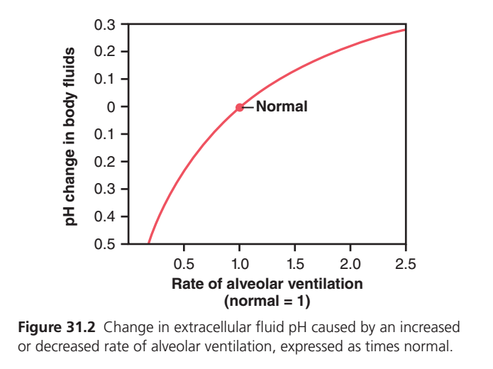
Figure 31.2: Effect of Alveolar Ventilation on Extracellular Fluid pH
- (Figure 31.2) shows how changes in alveolar ventilation affect the pH of body fluids (extracellular fluid).
Understanding the Axes
X-Axis (Horizontal)
Rate of Alveolar Ventilation (Normal = 1)
This shows how fast a person is breathing compared with normal.
| Value | Meaning |
|---|---|
| 0.5 | Breathing at half the normal rate (Hypoventilation) |
| 1.0 | Normal breathing |
| 1.5 | Breathing 1.5 times faster than normal |
| 2.0 | Breathing 2 times faster than normal |
| 2.5 | Breathing 2.5 times faster than normal (Hyperventilation) |
- Moving to the right = Breathing faster
- Moving to the left = Breathing slower
Y-Axis (Vertical)
pH Change in Body Fluids
This axis shows how much the extracellular fluid pH changes from the normal value.
Upper Half (Positive Values)
- +0.1
- +0.2
- +0.3
These indicate that pH increases.
- Blood becomes more alkaline (basic).
Middle Point (0)
This is the Normal pH.
- Normal ventilation
- Normal CO₂
- Normal blood pH
This point is marked “Normal” on the graph.
Lower Half (Negative Values)
The graph labels them as 0.1, 0.2, 0.3, 0.4, 0.5 below zero.
These represent a decrease in pH.
- Blood becomes more acidic.
Understanding the Red Curve
The red curve shows the relationship between:
Breathing Rate → CO₂ Level → Blood pH
Normal Point (Ventilation = 1)
At ventilation = 1
The graph passes through the Normal point.
Meaning:
- Normal breathing
- Normal CO₂ removal
- Normal extracellular fluid pH
No pH change occurs.
Left Side of the Curve (Ventilation Less Than Normal)
Move left from the normal point.
Example:
Ventilation decreases from 1 → 0.5
This means:
- Breathing becomes slower.
- Less CO₂ is removed by the lungs.
- CO₂ accumulates in the blood.
- More carbonic acid (H₂CO₃) forms.
- H⁺ concentration increases.
- Blood pH decreases.
Result:
Blood becomes more acidic.
The graph moves downward.
Right Side of the Curve (Ventilation Greater Than Normal)
Move right from the normal point.
Example:
Ventilation increases from 1 → 2
This means:
- Breathing becomes faster.
- More CO₂ is removed by the lungs.
- Blood CO₂ decreases.
- Carbonic acid (H₂CO₃) decreases.
- H⁺ concentration decreases.
- Blood pH increases.
Result:
Blood becomes more alkaline (basic).
The graph moves upward.
Why is the Curve Curved Instead of a Straight Line?
Notice:
Near the normal point, the curve is steeper.
At very high ventilation, the curve becomes flatter.
This means:
- Around normal breathing, small changes in ventilation produce noticeable changes in pH.
- At very high ventilation rates, further increases in breathing cause only smaller additional increases in pH.
Understanding the Entire Curve
Far Left (Very Slow Breathing)
- Very low ventilation
- CO₂ retained
- H₂CO₃ increases
- H⁺ increases
- pH falls markedly
Result: Severe acidosis.
Middle (Normal)
- Normal ventilation
- Normal CO₂
- Normal H⁺
- Normal pH
Far Right (Very Fast Breathing)
- Very high ventilation
- Excess CO₂ removed
- H₂CO₃ decreases
- H⁺ decreases
- pH rises
Result: Alkalosis.
Easy Concept
Think of the lungs as a CO₂ remover.
If you breathe slowly
➡ CO₂ stays in the blood.
➡ More acid forms.
➡ pH falls.
If you breathe faster
➡ CO₂ leaves the blood.
➡ Less acid remains.
➡ pH rises.
One-Line Memory Trick
Slow breathing → ↑ CO₂ → ↑ H⁺ → ↓ pH → Acidosis
Fast breathing → ↓ CO₂ → ↓ H⁺ → ↑ pH → Alkalosis
KEY CONCEPT
- Figure 31.2 shows how alveolar ventilation changes extracellular fluid pH.
- Normal ventilation (1× normal) maintains normal blood pH.
- Decreased ventilation (hypoventilation) causes CO₂ retention, H⁺ increases, and pH decreases (acidosis).
- Increased ventilation (hyperventilation) causes more CO₂ removal, H⁺ decreases, and pH increases (alkalosis).
- The lungs regulate blood pH by controlling the amount of CO₂ removed from the body.
Increased H⁺ Concentration Stimulates Alveolar Ventilation
Figure Mentioned: Fig. 31.3
- Alveolar ventilation not only changes H⁺ concentration.
- H⁺ concentration also controls the rate of alveolar ventilation.
- Fig. 31.3 shows the relationship between blood pH and alveolar ventilation.
- As pH decreases from the normal value of 7.4 to 7.0, alveolar ventilation increases 4 to 5 times normal.
Mathematical Values
Normal pH7.4
↓
Acidic pH7.0
↓
Alveolar ventilation4–5×Normal
Easy Concept
↓ pH (↑ H⁺)
↓
Respiratory center stimulated
↓
Alveolar ventilation increases
↓
4–5 × Normal
- When plasma pH rises above 7.4, alveolar ventilation decreases.
Easy Concept
↑ pH (↓ H⁺)
↓
Respiratory center depressed
↓
Ventilation decreases
- The change in ventilation for each unit change in pH is much greater when pH is low.
- The response is stronger when H⁺ concentration is high.
- The response is weaker when pH is high.
Easy Concept
Low pH
↓
Strong respiratory response
High pH
↓
Weak respiratory response
- Increased pH decreases alveolar ventilation.
- Decreased ventilation lowers the amount of oxygen entering the blood.
- Blood PO₂ decreases.
- Low PO₂ stimulates ventilation.
- Therefore, respiratory compensation for increased pH is limited.
- Respiratory compensation is much more effective during marked decreases in pH.
Easy Concept
↑ pH
↓
↓ Ventilation
↓
↓ Oxygen (PO₂)
↓
Oxygen receptors stimulated
↓
Ventilation increases again
↓
Compensation for alkalosis is limited
Feedback Control of H⁺ Concentration By the Respiratory System
- Increased H⁺ concentration stimulates respiration.
- Increased alveolar ventilation decreases H⁺ concentration.
- Therefore, the respiratory system works as a negative feedback controller of H⁺ concentration.
Easy Concept
↑ H⁺
↓
↑ Respiration
↓
↑ CO₂ Removal
↓
↓ CO₂
↓
↓ H₂CO₃
↓
↓ H⁺
↓
Back to Normal
- When H⁺ concentration rises above normal, the respiratory system is stimulated.
- Alveolar ventilation increases.
- Increased ventilation decreases extracellular PCO₂.
- Lower PCO₂ decreases H⁺ concentration.
- H⁺ concentration returns toward normal.
Easy Concept
↑ H⁺
↓
↑ Ventilation
↓
↓ PCO₂
↓
↓ H⁺
↓
Normal pH restored
- When H⁺ concentration falls below normal, the respiratory center is depressed.
- Alveolar ventilation decreases.
- Decreased ventilation increases PCO₂.
- Increased PCO₂ increases H⁺ concentration.
- H⁺ concentration returns toward normal.
Easy Concept
↓ H⁺
↓
↓ Ventilation
↓
↑ PCO₂
↓
↑ H⁺
↓
Normal pH restored
- Alkalosis depresses the respiratory centers.
- The respiratory response during alkalosis is weaker.
- The respiratory response during alkalosis is less predictable.
- Reduced alveolar ventilation causes hypoxemia.
- Hypoxemia activates oxygen-sensitive chemoreceptors.
- Oxygen-sensitive chemoreceptors stimulate ventilation.
- This limits respiratory compensation during metabolic alkalosis.
Easy Concept
Metabolic Alkalosis
↓
↓ Ventilation
↓
Hypoxemia
↓
Oxygen-sensitive chemoreceptors activated
↓
Ventilation increases
↓
Respiratory compensation is limited
KEY CONCEPT
- Figure Mentioned: Fig. 31.3
- Fig. 31.3 shows that decreasing pH increases alveolar ventilation.
- pH 7.4 → 7.0 increases ventilation to about 4–5 times normal.
- ↑ H⁺ → ↑ Ventilation → ↓ PCO₂ → ↓ H₂CO₃ → ↓ H⁺.
- ↓ H⁺ → ↓ Ventilation → ↑ PCO₂ → ↑ H₂CO₃ → ↑ H⁺.
- The respiratory system acts as a negative feedback mechanism to maintain normal H⁺ concentration.
- Respiratory compensation is stronger during acidosis than during alkalosis.
- During alkalosis, hypoxemia stimulates oxygen-sensitive chemoreceptors, limiting the decrease in ventilation.
Mathematical/Biochemical Equations Solved
- Normal pH → Acidic pH
7.4→7.0
- Ventilation Response
Alveolar Ventilation=4–5×Normal
- Acidosis Feedback
↑H+→↑Ventilation→↓PCO2→↓H2CO3→↓H+
- Alkalosis Feedback
↓H+→↓Ventilation→↑PCO2→↑H2CO3→↑H+
- Respiratory Compensation During Alkalosis
↓Ventilation→↓PO2→Hypoxemia→Chemoreceptor Stimulation→↑Ventilation
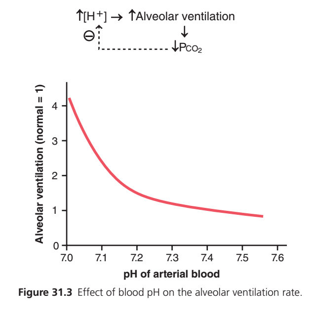
Figure 31.3: Effect of Blood pH on the Alveolar Ventilation Rate
- (Figure 31.3) shows how changes in arterial blood pH affect the rate of alveolar ventilation (breathing).
Understanding the Axes
X-Axis (Horizontal)
pH of Arterial Blood
This shows how acidic or alkaline the blood is.
| pH | Meaning |
|---|---|
| 7.0 | Very acidic |
| 7.1 | Acidic |
| 7.2 | Mildly acidic |
| 7.3 | Slightly acidic |
| 7.4 | Normal blood pH |
| 7.5 | Alkaline |
| 7.6 | More alkaline |
- Moving left = Blood becomes more acidic (↑ H⁺).
- Moving right = Blood becomes more alkaline (↓ H⁺).
Y-Axis (Vertical)
Alveolar Ventilation (Normal = 1)
This shows the breathing rate compared with normal.
| Value | Meaning |
|---|---|
| 1 | Normal breathing |
| 2 | 2 times normal breathing |
| 3 | 3 times normal breathing |
| 4 | 4 times normal breathing |
- Moving up = Faster breathing.
- Moving down = Slower breathing.
Understanding the Red Curve
The red curve shows the relationship between:
Blood pH → Breathing Rate
Left Side of the Curve (Low pH)
Look at the left end of the graph.
Blood pH is about 7.0.
This means:
- Blood is very acidic.
- H⁺ concentration is high.
The body responds by:
- Increasing alveolar ventilation to about 4 times normal.
Why?
- Faster breathing removes more CO₂.
- Less CO₂ means less carbonic acid (H₂CO₃).
- Less H₂CO₃ means fewer H⁺ ions.
- Blood pH increases toward normal.
Result: Severe acidosis causes a marked increase in breathing.
Middle of the Curve
As blood pH increases from 7.1 → 7.3:
- Blood becomes less acidic.
- H⁺ concentration decreases.
- The need for rapid breathing decreases.
Therefore:
- Alveolar ventilation gradually decreases.
Normal Point (pH ≈ 7.4)
At about pH 7.4:
- Blood pH is normal.
- H⁺ concentration is normal.
- Alveolar ventilation is approximately 1× normal.
This is the normal operating point.
Right Side of the Curve (High pH)
Move toward pH 7.5–7.6.
Now:
- Blood becomes alkaline.
- H⁺ concentration decreases.
The respiratory center receives less stimulation.
Therefore:
- Breathing becomes slower than normal.
Less CO₂ is removed.
CO₂ begins to accumulate.
More H₂CO₃ forms.
More H⁺ is produced.
Blood pH moves back toward normal.
Why is the Curve Curved?
Notice:
The curve is very steep at low pH.
This means:
A small fall in pH causes a large increase in breathing.
As pH becomes normal or alkaline,
the curve becomes flatter.
This means:
Further increases in pH produce only small decreases in breathing.
Understanding the Diagram Above the Graph
The small diagram summarizes the body’s negative feedback response.
Step 1
↑ H⁺
Means:
Hydrogen ion concentration increases.
Blood becomes acidic.
↓
Step 2
↑ Alveolar Ventilation
The respiratory center stimulates faster breathing.
↓
Step 3
↓ PCO₂
More CO₂ is exhaled.
Blood CO₂ falls.
↓
Step 4
Less CO₂ produces less carbonic acid (H₂CO₃).
↓
Step 5
H⁺ concentration decreases.
Blood pH returns toward normal.
Meaning of the Dotted Arrow and (⊖)
The dotted arrow points from ↓ PCO₂ back toward ↑ H⁺.
The ⊖ (negative sign) represents negative feedback.
Meaning:
- An increase in H⁺ starts the response.
- The lungs increase ventilation.
- CO₂ decreases.
- H⁺ decreases.
Therefore, the original increase in H⁺ is opposed (reduced).
This is a negative feedback mechanism that stabilizes blood pH.
Easy Concept
Imagine the lungs as an automatic CO₂ exhaust fan.
When blood becomes acidic
- H⁺ increases.
- The fan works faster.
- More CO₂ is blown out.
- Acid decreases.
- Blood pH returns toward normal.
When blood becomes alkaline
- H⁺ decreases.
- The fan slows down.
- Less CO₂ is removed.
- CO₂ builds up.
- More H⁺ forms.
- Blood pH falls back toward normal.
One-Line Memory Trick
↓ pH (↑ H⁺) → ↑ Breathing → ↓ CO₂ → ↓ H⁺ → pH returns toward normal
↑ pH (↓ H⁺) → ↓ Breathing → ↑ CO₂ → ↑ H⁺ → pH returns toward normal
KEY CONCEPT
- Figure 31.3 shows the effect of arterial blood pH on alveolar ventilation.
- Low blood pH (high H⁺) strongly stimulates alveolar ventilation.
- High blood pH (low H⁺) reduces alveolar ventilation.
- Increased breathing removes CO₂, reducing H₂CO₃ and H⁺, which raises blood pH.
- Decreased breathing retains CO₂, increasing H₂CO₃ and H⁺, which lowers blood pH.
- This is a negative feedback mechanism that helps maintain normal blood pH.
Efficiency of Respiratory Control of H⁺ Concentration
- The respiratory system cannot completely restore H⁺ concentration to normal when the disturbance is caused outside the respiratory system.
- Respiratory control is usually 50% to 75% effective.
- This corresponds to a feedback gain of 1 to 3 during metabolic acidosis.
Mathematical Values
Respiratory effectiveness:50% to 75%
Feedback gain:1 to 3
Easy Concept
Respiratory compensation
↓
Does not correct 100%
↓
Corrects about
50–75%
- If acid is suddenly added to the extracellular fluid, pH decreases.
- The pH may fall from 7.4 to 7.0.
Mathematical Solution
Initial pH7.4
↓
After acid is added7.0
- The respiratory system increases ventilation.
- This response raises the pH to about 7.2–7.3.
Mathematical Solution
After respiratory compensation7.2 to 7.3
Easy Concept
Normal pH7.4
↓
Acid Added
↓7.0
↓
Respiratory Compensation
↓7.2–7.3
Not completely back to 7.4
- Respiratory compensation occurs within 3 to 12 minutes.
Mathematical Values
3–12 minutes
- Respiratory responses to metabolic alkalosis are rapid.
- Respiratory compensation during metabolic alkalosis is limited.
- This limitation is caused by hypoxemia due to reduced alveolar ventilation.
Easy Concept
Metabolic Alkalosis
↓
↓ Ventilation
↓
Hypoxemia
↓
Compensation Limited
Buffering Power of the Respiratory System
- Respiratory regulation acts as a physiological buffer system.
- It responds rapidly.
- It prevents large changes in H⁺ concentration.
- It provides protection until the kidneys respond.
- The kidneys respond more slowly.
- The kidneys remove the acid–base imbalance.
- The buffering power of the respiratory system is 1 to 2 times greater than all extracellular chemical buffers combined.
Mathematical Values
1–2×
Easy Concept
Respiratory Buffer
↓
1–2 times stronger
↓
Than all chemical buffers combined
- The respiratory system buffers 1–2 times more acid or base than chemical buffers.
Impairment of Lung Function Can Cause Respiratory Acidosis
- Normal respiration helps buffer changes in H⁺ concentration.
- Abnormal respiration can also change H⁺ concentration.
- Severe emphysema is an example of impaired lung function.
- Impaired lungs cannot remove CO₂ effectively.
- CO₂ accumulates in the extracellular fluid.
- Increased CO₂ causes respiratory acidosis.
Easy Concept
Lung Disease
↓
↓ CO₂ Removal
↓
↑ CO₂
↓
↑ H₂CO₃
↓
↑ H⁺
↓
Respiratory Acidosis
- Lung disease also reduces the body’s ability to compensate for metabolic acidosis.
- Normally, metabolic acidosis increases ventilation.
- Increased ventilation lowers PCO₂.
- In lung disease, this compensatory decrease in PCO₂ is reduced.
Easy Concept
Metabolic Acidosis
↓
Should increase ventilation
↓
Lung disease prevents adequate ventilation
↓
PCO₂ remains high
↓
Poor compensation
- After initial chemical buffering, the kidneys become the only remaining physiological mechanism.
- The kidneys gradually return the pH toward normal.
Easy Concept
Chemical Buffers
↓
Respiratory Compensation Fails
↓
Kidneys become the only long-term regulator
↓
pH gradually returns toward normal
KEY CONCEPT
- Respiratory compensation is 50–75% effective.
- Feedback gain = 1–3 during metabolic acidosis.
- Acid can lower pH from 7.4 → 7.0.
- Respiratory compensation raises pH to about 7.2–7.3 within 3–12 minutes.
- Respiratory compensation is limited during metabolic alkalosis because of hypoxemia.
- The respiratory system has 1–2 times greater buffering power than all extracellular chemical buffers combined.
- Lung diseases (e.g., emphysema) reduce CO₂ elimination.
- CO₂ retention → ↑ H₂CO₃ → ↑ H⁺ → Respiratory acidosis.
- When respiratory compensation is impaired, the kidneys become the main mechanism for restoring normal pH.
Mathematical/Biochemical Equations Solved
- Respiratory effectiveness
50%−75%
- Feedback gain
1−3
- Respiratory compensation
7.4→7.0→7.2−7.3
- Time required
3−12 minutes
- Buffering power
1−2×
- Respiratory acidosis pathway
↓Lung Function→↑CO2→↑H2CO3→↑H+→Respiratory Acidosis
MADE BY SELF LEARNING DR SHEEN
