


- Blood volume and extracellular fluid volume are usually controlled in parallel.
- Ingested fluid is first absorbed from the gastrointestinal tract into the blood.
- The absorbed fluid is rapidly distributed between the plasma and interstitial spaces.
- Therefore, blood volume and extracellular fluid volume are usually regulated simultaneously.
- In some situations, the distribution of extracellular fluid between the blood and interstitial spaces can change greatly.
- The main factors that cause fluid accumulation in the interstitial spaces are:
- Increased capillary hydrostatic pressure.
- Decreased plasma colloid osmotic pressure.
- Increased capillary permeability.
- Obstruction of lymphatic vessels.
- In all of these conditions, a larger proportion of extracellular fluid moves into the interstitial spaces.
- Interstitial fluid volume is equal to extracellular fluid volume minus blood volume.
- Under normal conditions, blood volume increases as extracellular fluid volume increases.
- When small amounts of fluid accumulate because of excessive fluid intake or decreased renal fluid output, about 20% to 30% of the added fluid remains in the blood.
- This fluid increases blood volume.
- The remaining fluid enters the interstitial spaces.
- When extracellular fluid volume increases more than 30% to 50% above normal, almost all additional fluid enters the interstitial spaces.
- Very little of the additional fluid remains in the blood.
- Interstitial fluid pressure is normally negative.
- When interstitial fluid pressure becomes positive, the interstitial spaces become highly compliant.
- Once the tissues become highly compliant, large amounts of fluid enter the tissues.
- Interstitial fluid pressure then increases only slightly despite large fluid accumulation.
- The safety factor against edema normally depends on rising interstitial fluid pressure.
- Rising interstitial fluid pressure opposes further fluid accumulation in the tissues.
- This safety factor is lost when the tissues become highly compliant.
- Under normal conditions, the interstitial spaces act as an overflow reservoir for excess fluid.
- Interstitial fluid volume can increase by 10 to 30 liters.
- This accumulation of fluid causes edema.
- The interstitial spaces also function as an overflow release valve for the circulation.
- This protects the cardiovascular system from dangerous fluid overload.
- This protection helps prevent pulmonary edema.
- This protection helps prevent cardiac failure.
- Extracellular fluid volume and blood volume are often controlled simultaneously.
- The amount of fluid distributed between blood and interstitial spaces depends on the physical properties of the circulation.
- The amount of fluid distributed between blood and interstitial spaces depends on the physical properties of the interstitial spaces.
- The amount of fluid distributed between blood and interstitial spaces depends on fluid exchange across capillary membranes.
KEY CONCEPT
- Blood volume and extracellular fluid volume are usually regulated together.
- Excess fluid is distributed between blood and interstitial spaces.
- Mild fluid accumulation increases both blood volume and interstitial fluid volume.
- Severe extracellular fluid expansion causes most excess fluid to enter the interstitial spaces.
- The interstitial spaces act as an overflow reservoir for excess fluid.
- This helps protect the cardiovascular system from fluid overload, pulmonary edema, and cardiac failure.
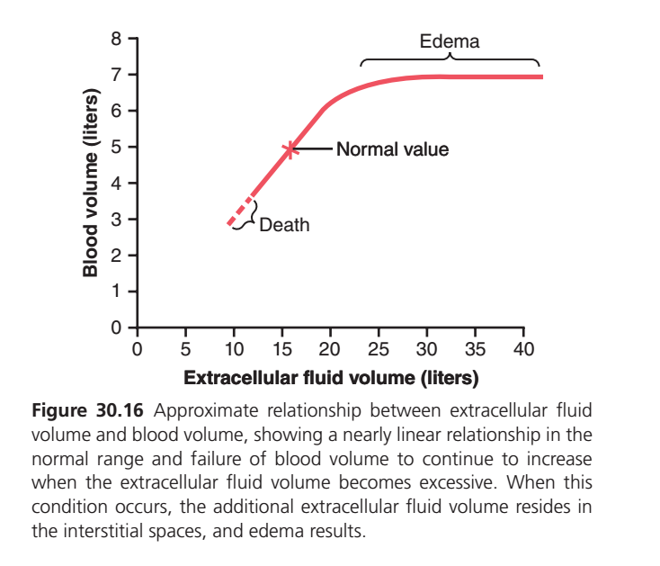
FIGURE 30.16: RELATIONSHIP BETWEEN EXTRACELLULAR FLUID (ECF) VOLUME AND BLOOD VOLUME
WHAT IS THIS GRAPH SHOWING?
This graph shows:
How changes in extracellular fluid (ECF) volume affect blood volume.
The main message is:
👉 In the normal range, blood volume and ECF volume increase together.
👉 But when ECF becomes excessively high, blood volume stops increasing and the extra fluid accumulates in tissues, causing edema.
UNDERSTANDING THE AXES
X-AXIS
Extracellular Fluid Volume (Liters)
This includes:
- Plasma fluid
- Interstitial fluid
Moving to the right means:
➡️ More extracellular fluid in the body.
Y-AXIS
Blood Volume (Liters)
This is the total volume of blood in the circulation.
THE RED CURVE
This red curve shows how blood volume changes as extracellular fluid volume changes.
PART 1: LEFT SIDE OF THE CURVE (DEATH ZONE)
ECF Volume ≈ 10 Liters
Blood volume is very low.
Approximately:
Blood Volume ≈ 3 Liters
This region is labeled:
DEATH
Why?
Very low extracellular fluid volume causes:
⬇️
Low blood volume
⬇️
Severe fall in blood pressure
⬇️
Poor tissue perfusion
⬇️
Death
PART 2: NORMAL LINEAR REGION
ECF Volume Increases
10 L → 15 L → 20 L
Blood volume also increases.
The relationship is almost a straight line.
What Does This Mean?
When extracellular fluid increases normally:
Some fluid enters the blood vessels
Therefore:
Blood volume increases almost proportionally.
NORMAL VALUE POINT ⭐
The star marks the:
Normal Operating Point
Approximately:
- ECF Volume = 15 L
- Blood Volume = 5 L
This is the normal physiological condition.
Easy Memory
Normal ECF = 15 L
Normal Blood Volume = 5 L
PART 3: CURVE STARTS FLATTENING
As ECF volume rises above normal:
Blood volume still increases
but
The increase becomes smaller and smaller.
Why?
The circulation can hold only a limited amount of extra fluid.
More fluid begins moving into tissues instead of remaining in blood vessels.
PART 4: EDEMA REGION
ECF Volume > 25–30 Liters
The curve becomes almost flat.
Notice:
Blood volume remains around 7 Liters
even though ECF volume continues increasing.
What Does a Flat Curve Mean?
Large increases in extracellular fluid
❌ Do NOT cause large increases in blood volume.
Instead:
✅ Extra fluid accumulates in interstitial spaces.WHY DOES EDEMA OCCUR?
When blood vessels are already relatively full:
Additional extracellular fluid cannot stay inside the circulation.
Therefore:
Excess Fluid
⬇️
Moves into Tissues
⬇️
Interstitial Fluid Increases
⬇️
Edema Develops
EASY CONCEPT
Imagine a water tank connected to a sponge.
First
Adding water fills the tank.
(Blood volume increases)
Later
The tank becomes nearly full.
Extra water starts soaking into the sponge.
(Interstitial fluid increases)
Result
Edema develops.
IMPORTANT MESSAGE OF THE GRAPH
Early Increase in ECF
⬇️
Blood volume increases.
Excessive Increase in ECF
⬇️
Blood volume increases very little.
⬇️
Most extra fluid enters tissues.
⬇️
Edema develops.
ONE-LINE INTERPRETATION OF EACH PART
Left Side
Very low ECF → Very low blood volume → Death.
Middle Part
ECF ↑ → Blood volume ↑ (nearly linear relationship).
Upper Flat Part
ECF ↑↑ → Blood volume changes little → Edema develops.
HIGH-YIELD EXAM POINT
Why does blood volume stop increasing despite continued ECF expansion?
✅ Because excess extracellular fluid shifts into the interstitial spaces, producing edema rather than further expanding blood volume.EY CONCEPT
✅ Figure 30.16 shows the relationship between extracellular fluid volume and blood volume.
✅ In the normal range, blood volume increases almost linearly with extracellular fluid volume.
✅ The normal operating point is approximately 15 L ECF volume and 5 L blood volume.
✅ At very low ECF volumes, blood volume falls to life-threatening levels.
✅ When ECF volume becomes excessively high, blood volume plateaus.
✅ Additional extracellular fluid accumulates in interstitial spaces rather than in blood vessels.
✅ This excess interstitial fluid causes edema.
Memory Formula
Normal ECF Expansion → ↑ Blood Volume
Excessive ECF Expansion → ↑ Interstitial Fluid → Edema ✔️
Made by self learning CEO and founder Dr sheen
