


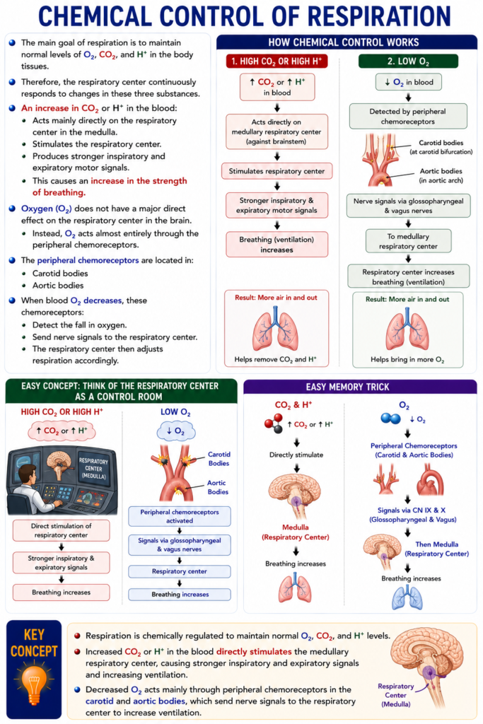
- The main goal of respiration is to maintain normal levels of O₂, CO₂, and H⁺ in the body tissues.
- Therefore, the respiratory center continuously responds to changes in these three substances.
- An increase in CO₂ or H⁺ in the blood:
- Acts mainly directly on the respiratory center in the medulla.
- Stimulates the respiratory center.
- Produces stronger inspiratory and expiratory motor signals.
- This causes an increase in the strength of breathing.
- Oxygen (O₂) does not have a major direct effect on the respiratory center in the brain.
- Instead, O₂ acts almost entirely through the peripheral chemoreceptors.
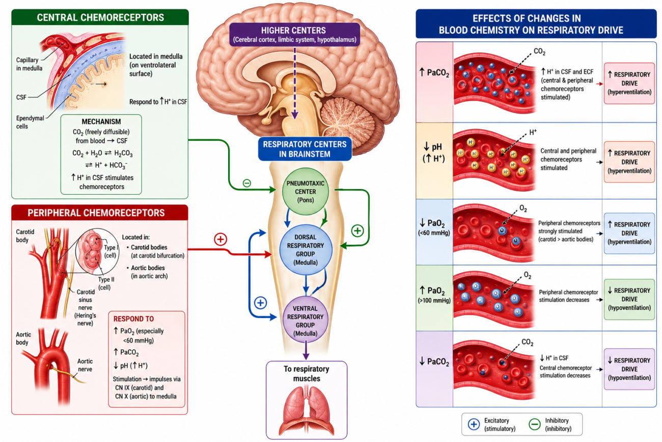
- The peripheral chemoreceptors are located in:
- Carotid bodies
- Aortic bodies
- When blood O₂ decreases, these chemoreceptors:
- Detect the fall in oxygen.
- Send nerve signals to the respiratory center.
- The respiratory center then adjusts respiration accordingly.
Easy Concept
Think of the respiratory center as a control room.
High CO₂ or High H⁺
↑ CO₂ or ↑ H⁺
        ↓
Medullary Respiratory Center
        ↓
Stronger inspiratory & expiratory signals
        ↓
Breathing increases
Low O₂
↓ O₂
      ↓
Carotid & Aortic Bodies
      ↓
Peripheral Chemoreceptors
      ↓
Respiratory Center
      ↓
Breathing increases
Easy Memory Trick
CO₂ & H⁺
↓↓
Directly stimulate
Medulla
O₂
↓↓
Peripheral Chemoreceptors
(Carotid & Aortic Bodies)
↓↓
Then MedullaKEY CONCEPT
- Respiration is chemically regulated to maintain normal O₂, CO₂, and H⁺ levels. Increased CO₂ or H⁺ directly stimulates the medullary respiratory center to increase breathing, whereas decreased O₂ acts mainly through the carotid and aortic body peripheral chemoreceptors, which send signals to the respiratory center to regulate ventilation.
DIRECT CONTROL OF RESPIRATORY CENTER ACTIVITY BY CO₂ AND H⁺
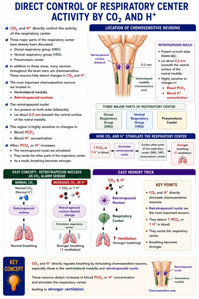
- CO₂ and H⁺ directly control the activity of the respiratory center.
- Three major parts of the respiratory center have already been discussed:
- Dorsal respiratory group (DRG)
- Ventral respiratory group (VRG)
- Pneumotaxic center
- In addition to these areas, many neurons throughout the brain stem are chemosensitive.
- These neurons help detect changes in CO₂ and H⁺.
- The most important chemosensitive neurons are located in:
- Ventrolateral medulla
- Retrotrapezoid nucleus
- The retrotrapezoid nuclei:
- Are present on both sides (bilaterally).
- Lie about 0.2 mm beneath the ventral surface of the rostral medulla.
- This region is highly sensitive to changes in:
- Blood PCO₂
- Blood H⁺ concentration
- When PCO₂ or H⁺ increases:
- The retrotrapezoid nuclei are stimulated.
- They excite the other parts of the respiratory center.
- As a result, breathing becomes stronger.
Easy Concept
Think of the retrotrapezoid nucleus as the CO₂ alarm sensor.
Normal CO₂
Normal CO₂
      ↓
Retrotrapezoid nucleus
      ↓
Normal breathing
Increased CO₂ or H⁺
↑ CO₂ or ↑ H⁺
        ↓
Retrotrapezoid nucleus detects change
        ↓
Stimulates respiratory center
        ↓
Stronger breathing
Easy Memory Trick
CO₂ & H⁺
        ↓
Retrotrapezoid Nucleus
        ↓
Respiratory Center
        ↓
↑ Ventilation
KEY CONCEPT
- CO₂ and H⁺ directly regulate breathing by stimulating chemosensitive neurons, especially those in the ventrolateral medulla and retrotrapezoid nuclei. These neurons detect increases in blood PCO₂ or H⁺ concentration and stimulate the respiratory center, leading to stronger ventilation.
EXCITATION OF THE CHEMOSENSITIVE NEURONS BY H⁺
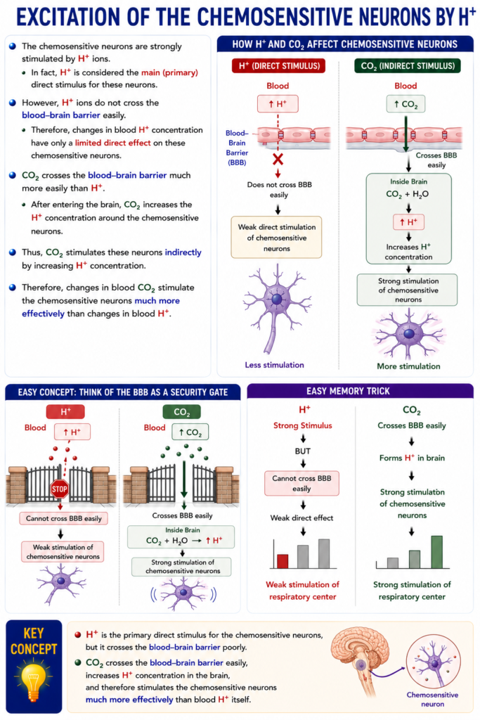
- The chemosensitive neurons are strongly stimulated by H⁺ ions.
- In fact, H⁺ is considered the main (primary) direct stimulus for these neurons.
- However, H⁺ ions do not cross the blood–brain barrier easily.
- Therefore, changes in blood H⁺ concentration have only a limited direct effect on these chemosensitive neurons.
- CO₂ crosses the blood–brain barrier much more easily than H⁺.
- After entering the brain, CO₂ increases the H⁺ concentration around the chemosensitive neurons.
- Thus, CO₂ stimulates these neurons indirectly by increasing H⁺ concentration.
- Therefore, changes in blood CO₂ stimulate the chemosensitive neurons much more effectively than changes in blood H⁺.
Easy Concept
Think of the blood–brain barrier (BBB) as a security gate.
H⁺
Blood

↑ H⁺
   │
   ✖ Cannot cross BBB easily
   │
Weak stimulation of
chemosensitive neurons
CO₂
Blood

↑ CO₂
   │
   ✔ Crosses BBB easily
   │
Inside Brain
CO₂ + H₂O
      ↓
H⁺ ↑
      ↓
Strong stimulation of
chemosensitive neurons
Easy Memory Trick
H⁺
↓↓
Strong Stimulus

BUT

Cannot cross BBB easily
↓↓
Weak direct effect
CO₂
↓↓
Crosses BBB
↓↓
Forms H⁺
↓↓
Strong respiratory stimulationKEY CONCEPT
- H⁺ is the primary direct stimulus for the chemosensitive neurons, but it crosses the blood–brain barrier poorly. In contrast, CO₂ crosses the blood–brain barrier easily, increases H⁺ concentration in the brain, and therefore stimulates the chemosensitive neurons much more effectively than blood H⁺ itself.
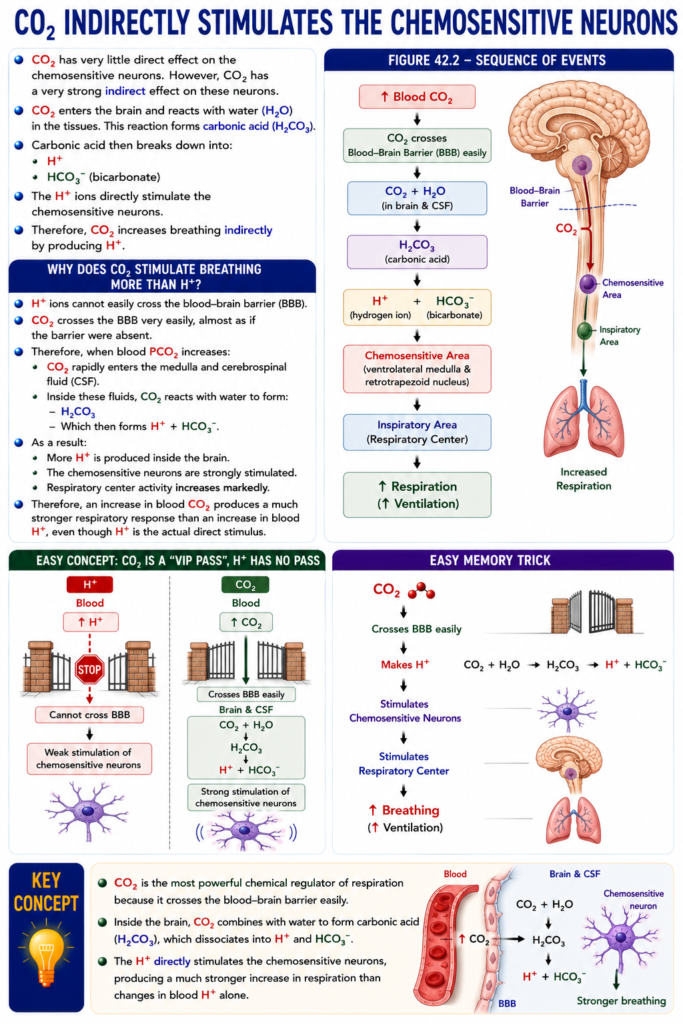
CO₂ INDIRECTLY STIMULATES THE CHEMOSENSITIVE NEURONS
- CO₂ has very little direct effect on the chemosensitive neurons.
- However, CO₂ has a very strong indirect effect on these neurons.
- CO₂ enters the brain and reacts with water (H₂O) in the tissues.
- This reaction forms carbonic acid (H₂CO₃).
- Carbonic acid then breaks down into:
- H⁺
- HCO₃⁻ (bicarbonate)
- The H⁺ ions directly stimulate the chemosensitive neurons.
- Therefore, CO₂ increases breathing indirectly by producing H⁺.
- Figure 42.2 illustrates these reactions.
Why Does CO₂ Stimulate Breathing More Than H⁺?
- H⁺ ions cannot easily cross the blood–brain barrier (BBB).
- CO₂ crosses the BBB very easily, almost as if the barrier were absent.
- Therefore, when blood PCO₂ increases:
- CO₂ rapidly enters the medulla and cerebrospinal fluid (CSF).
- Inside these fluids, CO₂ reacts with water to form:
- H₂CO₃
- Which then forms H⁺ + HCO₃⁻.
- As a result:
- More H⁺ is produced inside the brain.
- The chemosensitive neurons are strongly stimulated.
- Respiratory center activity increases markedly.
- Therefore, an increase in blood CO₂ produces a much stronger respiratory response than an increase in blood H⁺, even though H⁺ is the actual direct stimulus.
Figure 42.2
Shows the sequence:
↑ Blood CO₂
      ↓
CO₂ crosses Blood–Brain Barrier
      ↓
CO₂ + H₂O
      ↓
H₂CO₃
      ↓
H⁺ + HCO₃⁻
      ↓
Chemosensitive Area
      ↓
Inspiratory Area
      ↓
↑ Respiration
Easy Concept
Think of CO₂ as a “VIP pass” and H⁺ as a person without a pass.
H⁺
Blood

↑ H⁺
      │
      ✖ Cannot cross BBB
      │
Weak stimulation
CO₂
Blood

↑ CO₂
      │
      ✔ Crosses BBB easily
      │
Brain & CSF

CO₂ + H₂O
      ↓
H₂CO₃
      ↓
H⁺
      ↓
Strong stimulation of
chemosensitive neurons
Easy Memory Trick
CO₂
↓↓
Crosses BBB
↓↓
Makes H⁺
↓↓
Stimulates Respiratory Center
↓↓
↑ Breathing
KEY CONCEPT
- CO₂ is the most powerful chemical regulator of respiration because it crosses the blood–brain barrier easily. Inside the brain, CO₂ combines with water to form carbonic acid, which dissociates into H⁺ and HCO₃⁻. The H⁺ directly stimulates the chemosensitive neurons, producing a much stronger increase in respiration than changes in blood H⁺ alone.
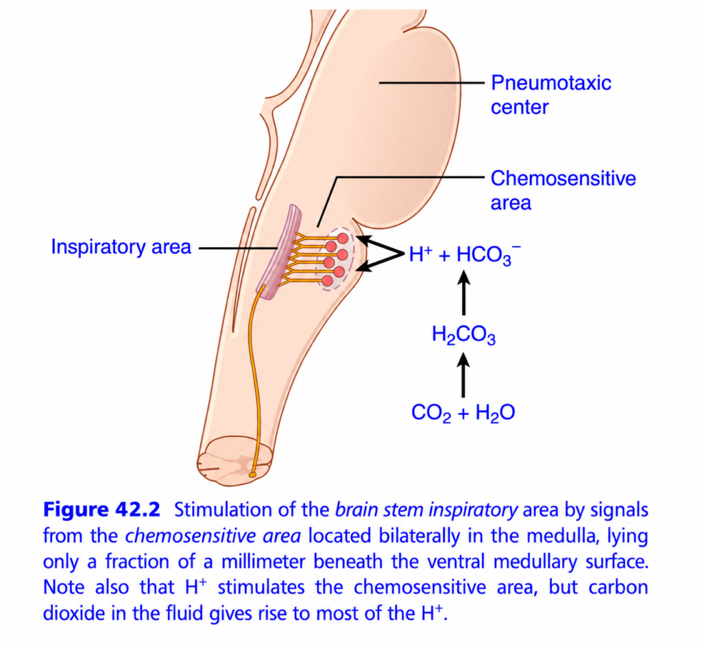
CO₂ Indirectly Stimulates the Chemosensitive Area (Ganong Fig. 42.2) – Easiest & Most Conceptual Explanation for MBBS Students
🎯 One-Line Concept
CO₂ is the most powerful chemical regulator of breathing because it indirectly increases H⁺ concentration in the brain, and H⁺ strongly stimulates the respiratory center to increase ventilation.
💡 Golden Rule
CO₂ is the messenger, but H⁺ is the real stimulator of the chemosensitive area.
The Big Picture
This figure explains how the brain knows when to breathe faster.
Imagine your brain has a “breathing control room.”
When CO₂ rises in the blood, this control room immediately tells your lungs:
“Breathe faster and deeper to remove the extra CO₂!”
Step-by-Step Story
Step 1. Body Cells Produce CO₂
Every cell produces CO₂ while making ATP.
Body Cells
      ↓
CO₂ produced
      ↓
Blood
Normally, this CO₂ is carried to the lungs and exhaled.
Step 2. Blood CO₂ Increases
Suppose you:
- Hold your breath
- Exercise
- Have lung disease
Then,
Blood CO₂ increases.
Normal CO₂
      ↓
High CO₂
Step 3. CO₂ Easily Crosses the Blood-Brain Barrier
This is the most important concept.
CO₂ crosses the blood-brain barrier very easily.
However,
H⁺ cannot cross easily.
This is why CO₂ is the main signal reaching the brain.
Blood
CO₂ ✔ Passes

H⁺ ✖ Cannot easily pass
Step 4. CO₂ Reacts with Water
Once CO₂ enters the brain tissue (or cerebrospinal fluid),
it reacts with water.
CO₂ + H₂O
      ↓
H₂CO₃
This forms
Carbonic Acid (H₂CO₃)
Step 5. Carbonic Acid Breaks Down
Carbonic acid is unstable.
It immediately dissociates into
H₂CO₃
      ↓
H⁺ + HCO₃⁻
Now,
H⁺ concentration increases.
Step 6. H⁺ Stimulates the Chemosensitive Area
This is the key point shown in the figure.
The chemosensitive area, located on the ventral surface of the medulla, is highly sensitive to H⁺.
↑ H⁺
      ↓
Chemosensitive Area Activated
Important
The chemosensitive area is not strongly stimulated by CO₂ directly.
Instead,
CO₂ works indirectly by generating H⁺.
Step 7. Chemosensitive Area Stimulates the Inspiratory Center
The activated chemosensitive area sends signals to the
Inspiratory Area (Dorsal Respiratory Group)
Chemosensitive Area
         ↓
Inspiratory Center
The inspiratory center then sends nerve impulses to:
- Diaphragm
- External intercostal muscles
Step 8. Breathing Increases
As a result,
- Respiratory rate increases.
- Depth of breathing increases.
More Breathing
       ↓
More CO₂ removed
Step 9. CO₂ Returns to Normal
The extra breathing removes CO₂ from the blood.
CO₂ ↓
      ↓
H⁺ ↓
      ↓
Respiratory center returns to normal
This is an example of negative feedback.
Complete Flow Chart
Body Cells
      │
Produce CO₂
      │
      ▼
Blood CO₂ rises
      │
      ▼
CO₂ crosses Blood-Brain Barrier
      │
      ▼
CO₂ + H₂O
      │
      ▼
H₂CO₃
      │
      ▼
H⁺ + HCO₃⁻
      │
      ▼
Chemosensitive Area
      │
      ▼
Inspiratory Center
      │
      ▼
Diaphragm Contracts
      │
      ▼
Breathing Increases
      │
      ▼
CO₂ Exhaled
      │
      ▼
CO₂ Returns to Normal
Why Doesn’t H⁺ Stimulate Directly from Blood?
This is a favorite MBBS viva question.
Answer
Hydrogen ions cannot easily cross the blood-brain barrier.
CO₂ crosses easily.
Once inside the brain,
CO₂ is converted into H⁺.
Therefore,
CO₂ indirectly stimulates the respiratory center.
Everyday Analogy
Imagine a house with a smoke detector.
🔥 CO₂ = Smoke
🚨 H⁺ = Alarm Sound
🧠 Brain = Fire Station
The smoke itself doesn’t ring the alarm.
Instead,
Smoke activates the detector,
which produces the alarm sound.
Similarly,
CO₂
      ↓
Produces H⁺
      ↓
H⁺ activates the brain
Structures in the Figure
1. Chemosensitive Area
📍 Located on the ventrolateral surface of the medulla.
Function
Detects increased H⁺ in the brain’s extracellular fluid/CSF and stimulates breathing.
2. Inspiratory Area
Located in the medulla.
Function
Generates the basic rhythm of inspiration and activates the respiratory muscles.3. Pneumotaxic Center
Located in the pons.
Function
Limits (switches off) inspiration, helping regulate the rate and depth of breathing.
Why Is CO₂ the Strongest Stimulus for Breathing?
Because:
- ✅ CO₂ crosses the blood-brain barrier rapidly.
- ✅ It quickly forms H⁺ in the brain.
- ✅ H⁺ powerfully stimulates the chemosensitive area.
- ✅ Ventilation increases within seconds.
This makes CO₂ the most important short-term regulator of ventilation.
High-Yield Comparison
| CO₂ | H⁺ |
|---|---|
| Crosses the blood-brain barrier easily | Crosses poorly |
| Indirectly stimulates respiration | Directly stimulates central chemoreceptors once formed in the brain |
| Forms H₂CO₃ | Final stimulant of the chemosensitive area |
MBBS High-Yield Points
- ✅ Central chemoreceptors respond primarily to H⁺ in the brain extracellular fluid/CSF.
- ✅ CO₂ is the major regulator because it readily crosses the blood-brain barrier and generates H⁺.
- ✅ H⁺ in the blood has little direct effect on central chemoreceptors because it crosses the blood-brain barrier poorly.
- ✅ An increase in CO₂ leads to increased ventilation, which removes CO₂ and restores normal levels (negative feedback).
🌟 Super Memory Summary
↑ CO₂ in Blood
        │
        ▼
Crosses Blood-Brain Barrier
        │
        ▼
CO₂ + H₂O
        │
        ▼
H₂CO₃
        │
        ▼
H⁺ + HCO₃⁻
        │
        ▼
Chemosensitive Area
        │
        ▼
Inspiratory Center
        │
        ▼
↑ Rate & Depth of Breathing
        │
        ▼
↑ CO₂ Exhalation
        │
        ▼
CO₂ Normal
🧠 Easy Mnemonic
“CO₂ → H⁺ → Breathe”
- CO₂ rises
- H⁺ is produced
- Brain stimulates breathing
- Extra CO₂ is exhaled
⭐ Golden Rule
CO₂ has very little direct effect on the chemosensitive neurons. Its powerful effect on breathing is indirect: it crosses the blood-brain barrier, forms carbonic acid, increases H⁺ concentration, and the H⁺ then strongly stimulates the medullary chemosensitive area, which activates the inspiratory center to increase ventilation.
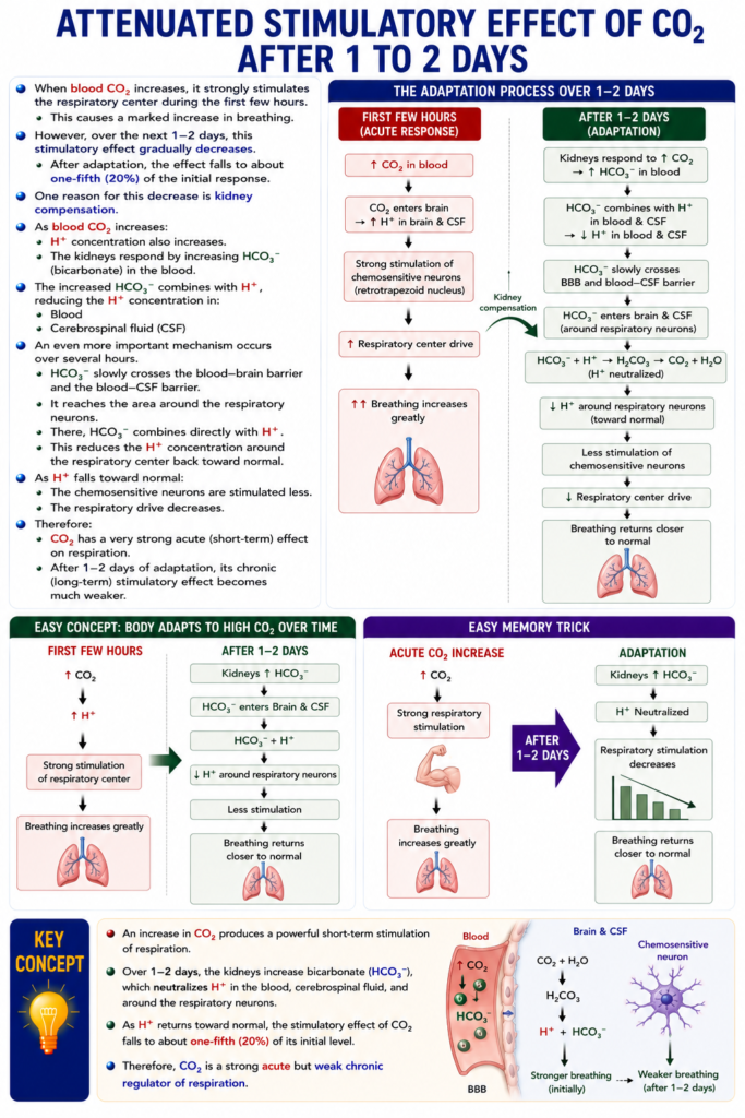
ATTENUATED ( Reduced) STIMULATORY EFFECT OF CO₂ AFTER 1 TO 2 DAYS
- When blood CO₂ increases, it strongly stimulates the respiratory center during the first few hours.
- This causes a marked increase in breathing.
- However, over the next 1–2 days, this stimulatory effect gradually decreases.
- After adaptation, the effect falls to about one-fifth (20%) of the initial response.
- One reason for this decrease is kidney compensation.
- As blood CO₂ increases:
- H⁺ concentration also increases.
- The kidneys respond by increasing HCO₃⁻ (bicarbonate) in the blood.
- The increased HCO₃⁻ combines with H⁺, reducing the H⁺ concentration in:
- Blood
- Cerebrospinal fluid (CSF)
- An even more important mechanism occurs over several hours.
- HCO₃⁻ slowly crosses the blood–brain barrier and the blood–CSF barrier.
- It reaches the area around the respiratory neurons.
- There, HCO₃⁻ combines directly with H⁺.
- This reduces the H⁺ concentration around the respiratory center back toward normal.
- As H⁺ falls toward normal:
- The chemosensitive neurons are stimulated less.
- The respiratory drive decreases.
- Therefore:
- CO₂ has a very strong acute (short-term) effect on respiration.
- After 1–2 days of adaptation, its chronic (long-term) stimulatory effect becomes much weaker.
Easy Concept
Think of the body as adjusting to high CO₂ over time.
First Few Hours
↑ CO₂
      ↓
↑ H⁺
      ↓
Strong stimulation
of respiratory center
      ↓
Breathing increases greatly
After 1–2 Days
Kidneys ↑ HCO₃⁻
      ↓
HCO₃⁻ enters Brain & CSF
      ↓
HCO₃⁻ + H⁺
      ↓
↓ H⁺ around respiratory neurons
      ↓
Less stimulation
      ↓
Breathing returns closer to normal
Easy Memory Trick
Acute CO₂ Increase
↓↓
Strong respiratory stimulation
After 1–2 Days
↓↓
Kidneys ↑ HCO₃⁻
↓↓
H⁺ Neutralized
↓↓
Respiratory stimulation decreasesKEY CONCEPT
- An increase in CO₂ produces a powerful short-term stimulation of respiration. Over 1–2 days, the kidneys increase bicarbonate (HCO₃⁻), which neutralizes H⁺ in the blood, cerebrospinal fluid, and around the respiratory neurons. As H⁺ returns toward normal, the stimulatory effect of CO₂ falls to about one-fifth of its initial level, making CO₂ a strong acute but weak chronic regulator of respiration.
QUANTITATIVE EFFECTS OF BLOOD PCO₂ AND H⁺ CONCENTRATION ON ALVEOLAR VENTILATION
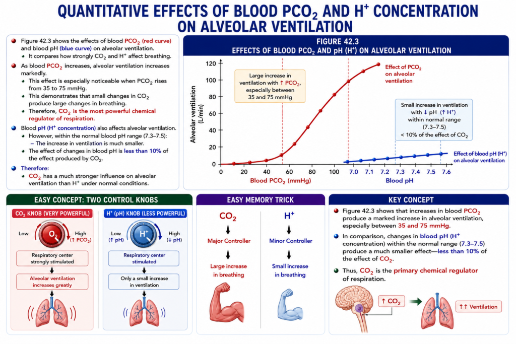
- Figure 42.3 shows the effects of blood PCO₂ (red curve) and blood pH (blue curve) on alveolar ventilation.
- It compares how strongly CO₂ and H⁺ affect breathing.
- As blood PCO₂ increases, alveolar ventilation increases markedly.
- This effect is especially noticeable when PCO₂ rises from 35 to 75 mmHg.
- This demonstrates that small changes in CO₂ produce large changes in breathing.
- Therefore, CO₂ is the most powerful chemical regulator of respiration.
- Blood pH (H⁺ concentration) also affects alveolar ventilation.
- However, within the normal blood pH range (7.3–7.5):
- The increase in ventilation is much smaller.
- The effect of changes in blood pH is less than 10% of the effect produced by CO₂.
- Therefore:
- CO₂ has a much stronger influence on alveolar ventilation than H⁺ under normal conditions.
Figure 42.3
Shows two curves:
- Red curve → Effect of PCO₂ on alveolar ventilation.
- Blue curve → Effect of blood pH (H⁺) on alveolar ventilation.
The figure demonstrates that:
- ↑ PCO₂ → Large increase in ventilation
- ↓ pH (↑ H⁺) → Small increase in ventilation
Easy Concept
Think of the respiratory center as having two control knobs.
CO₂ Knob (Very Powerful)
↑ PCO₂
      ↓
Respiratory center strongly stimulated
      ↓
Alveolar ventilation increases greatly
H⁺ (pH) Knob (Less Powerful)
↓ pH (↑ H⁺)
      ↓
Respiratory center stimulated
      ↓
Only a small increase in ventilation
Easy Memory Trick
CO₂
↓↓
Major Controller
↓↓
Large increase in breathing
H⁺
↓↓
Minor Controller
↓↓
Small increase in breathingKEY CONCEPT
- Figure 42.3 shows that increases in blood PCO₂ produce a marked increase in alveolar ventilation, especially between 35 and 75 mmHg. In comparison, changes in blood pH (H⁺ concentration) within the normal range (7.3–7.5) produce a much smaller effect—less than 10% of the effect of CO₂. Thus, CO₂ is the primary chemical regulator of respiration.
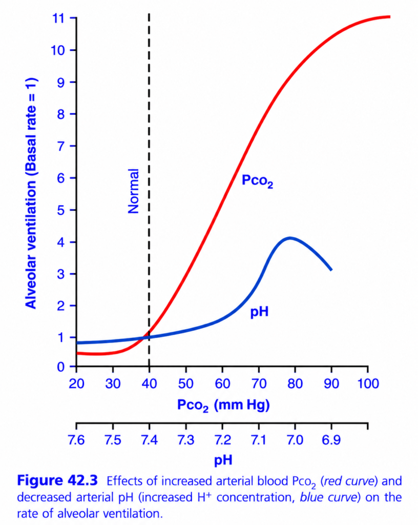
Quantitative Effects of Blood PCO₂ and H⁺ (pH) on Alveolar Ventilation (Ganong Fig. 42.3) – Easiest & Most Conceptual Explanation for MBBS Students
🎯 One-Line Concept
CO₂ is the strongest stimulus for breathing, while H⁺ (low pH) also increases breathing but has a weaker effect.
💡 Golden Rule
When CO₂ rises → breathing increases dramatically.
When pH falls (H⁺ rises) → breathing also increases, but less than CO₂.
First Understand the Graph
The graph compares two different stimuli that affect breathing.
- 🔴 Red Curve = Effect of CO₂ (PCO₂)
- 🔵 Blue Curve = Effect of H⁺ (Low pH)
Both increase alveolar ventilation, but CO₂ has a much stronger effect.
Step 1: Understand the Axes
X-Axis (Horizontal)
Shows:
- PCO₂ (20–100 mm Hg)
OR
- pH (7.6–6.9)
Moving to the right means:
- PCO₂ increases
- pH decreases (blood becomes more acidic)
Y-Axis (Vertical)
Shows
Alveolar Ventilation
Normal breathing is marked as
1 (Basal Rate)
Ventilation

11 ↑
10 │
 9 │
 8 │
 7 │
 6 │
 5 │
 4 │
 3 │
 2 │
 1 │ ← Normal breathing
 0 └───────────────→
Higher on the graph means
➡ Faster and deeper breathing.
The Dashed Vertical Line
The dashed line marks
Normal Arterial Values
PCO₂ = 40 mm Hg
pH = 7.4
At this point
Normal ventilation = 1
This is your resting breathing.
RED CURVE – Effect of CO₂ ⭐ (Most Important)
This is the most important part of the graph.
When CO₂ is Low (20–35 mm Hg)
Breathing is slightly reduced.
Why?
Because the body does not need to remove much CO₂.
Low CO₂
      ↓
Little stimulation
      ↓
Slow breathing
At Normal CO₂ (40 mm Hg)
Breathing remains normal.
PCO₂ = 40
      ↓
Normal ventilation
When CO₂ Rises Above 40 mm Hg
This is the key concept.
As CO₂ rises,
breathing increases very rapidly.
CO₂ ↑
     ↓
Brain stimulated
     ↓
Breathing ↑↑↑
The red curve becomes very steep.
Why?
Because
CO₂ crosses the blood-brain barrier,
forms H⁺,
stimulates the chemosensitive area,
which stimulates the inspiratory center.
Result
Very powerful increase in ventilation.
Example
Suppose
PCO₂ increases from
40 → 60 mm Hg
Breathing increases
approximately
5–6 times normal
This is why CO₂ is called
The Primary Chemical Stimulus for Breathing
At Very High CO₂ (>80–100 mm Hg)
The curve begins to flatten.
Why?
The respiratory center cannot continue increasing indefinitely.
Eventually,
very high CO₂ may even depress brain function.
BLUE CURVE – Effect of H⁺ (Low pH)
Now look at the blue curve.
Normal pH
pH = 7.4
Ventilation = Normal
Blood Becomes Acidic
When
pH falls
from
7.4 → 7.2 → 7.1
Hydrogen ions increase.
pH ↓
H⁺ ↑
Peripheral chemoreceptors (mainly carotid bodies) are stimulated, and ventilation increases.
Why Is the Increase Smaller?
Hydrogen ions cannot cross the blood-brain barrier easily.
Therefore,
they mainly stimulate the peripheral chemoreceptors, producing a weaker ventilatory response than CO₂.
At Severe Acidosis
The blue curve rises.
Breathing becomes faster.
But
Even at its highest,
the blue curve remains well below the red curve.
This shows
CO₂ is much more powerful than H⁺.
Why Does the Blue Curve Fall at Very Low pH?
Notice
At extremely low pH
(around 7.0)
the curve starts falling.
Why?
Because
Severe acidosis begins to depress the respiratory center and overall nervous system function.
So
Breathing cannot continue increasing forever.
Compare Both Curves
| CO₂ (Red Curve) | H⁺ / Low pH (Blue Curve) |
|---|---|
| Strongest stimulus | Weaker stimulus |
| Crosses blood-brain barrier easily | H⁺ crosses poorly |
| Stimulates central chemoreceptors indirectly (via H⁺ in CSF) | Mainly stimulates peripheral chemoreceptors |
| Ventilation increases dramatically | Ventilation increases moderately |
Easy Everyday Analogy
Imagine your house has two alarms.
🔴 CO₂ Alarm
Very sensitive.
Even a small increase
🚨 Alarm becomes loud immediately.
🔵 Acid Alarm
Works,
but is less sensitive.
It activates later
and not as strongly.
Flow Chart
CO₂ Pathway
CO₂ ↑
     │
Crosses Blood-Brain Barrier
     │
CO₂ + H₂O
     │
H₂CO₃
     │
H⁺ ↑
     │
Central Chemoreceptors
     │
Inspiratory Center
     │
Ventilation ↑↑↑
H⁺ Pathway
Blood H⁺ ↑
      │
Peripheral Chemoreceptors
(Carotid & Aortic Bodies)
      │
Respiratory Center
      │
Ventilation ↑
Why Is CO₂ More Powerful?
Because
✅ Crosses the blood-brain barrier easily.
✅ Produces H⁺ inside the brain.
✅ Strongly stimulates the medullary chemosensitive area.
Hydrogen ions in the blood cannot do this efficiently.
High-Yield Exam Points
Q1. What is the most powerful chemical stimulus for breathing?
✅ Increased arterial PCO₂
Q2. Why does CO₂ stimulate breathing more than H⁺?
✅ Because CO₂ easily crosses the blood-brain barrier and generates H⁺ in the cerebrospinal fluid, strongly stimulating the central chemoreceptors.
Q3. Which receptors respond mainly to metabolic acidosis?
✅ Peripheral chemoreceptors (carotid and aortic bodies)
Q4. Normal arterial values?
- PCO₂ = 40 mm Hg
- pH = 7.4
⭐ Super Memory Summary
               NORMAL
        PCO₂ = 40 mm Hg
           pH = 7.4
                │
                ▼
      Normal Ventilation (1)
                │
 ┌──────────────┴──────────────┐
 │                             │
 ▼                             ▼
CO₂ ↑                      H⁺ ↑ (pH ↓)
 │                             │
Crosses BBB               Peripheral
 │                         Chemoreceptors
 ▼                             ▼
Central Chemoreceptors     Respiratory Center
 │                             │
 ▼                             ▼
Ventilation ↑↑↑            Ventilation ↑
🧠 Easy Mnemonics
CO₂ Curve
“CO₂ Controls Breathing.”
- CO₂ ↑ → Ventilation ↑↑↑
pH Curve
“Acid Also Stimulates, but Less.”
- pH ↓ → Ventilation ↑
💎 Golden Rule
The red CO₂ curve is much steeper than the blue pH curve because carbon dioxide is the principal short-term regulator of ventilation. A small increase in arterial PCO₂ causes a large increase in alveolar ventilation, whereas an equivalent increase in H⁺ concentration produces a much smaller response, mainly through peripheral chemoreceptors.
CHANGES IN O₂ HAVE LITTLE DIRECT EFFECT ON CONTROL OF THE BRAIN RESPIRATORY CENTER
- Changes in blood O₂ have almost no direct effect on the respiratory center in the brain.
- Therefore, O₂ does not directly change the respiratory drive.
- However, O₂ does affect breathing indirectly through the peripheral chemoreceptors.
- The hemoglobin–oxygen buffer system helps deliver almost normal amounts of O₂ to the tissues, even when the pulmonary PO₂ changes widely.
- It continues to supply adequate oxygen when the pulmonary PO₂ ranges from about 60 mmHg to 1000 mmHg.
- Therefore, under normal conditions:
- Adequate tissue oxygen delivery is maintained.
- Even if lung ventilation decreases to less than half of normal.
- Or increases to 20 times or more than normal.
- This is not true for CO₂.
- Blood and tissue PCO₂ change directly with ventilation:
- ↓ Ventilation → ↑ PCO₂
- ↑ Ventilation → ↓ PCO₂
- Because CO₂ changes rapidly with ventilation, evolution has made CO₂ the main controller of respiration, not O₂.
- However, the body has a special protective mechanism for situations where oxygen becomes dangerously low.
- This mechanism is located in the peripheral chemoreceptors, which are outside the brain respiratory center.
- The peripheral chemoreceptors become strongly activated when arterial PO₂ falls below about 70 mmHg.
- They then send signals to the respiratory center to increase breathing.
Easy Concept
Think of CO₂ as the normal controller and O₂ as the emergency controller.
Normal Situation
Normal O₂
↓

Hemoglobin supplies enough O₂

↓

Respiratory center is NOT affected much
CO₂ Changes
↑ CO₂
↓

Respiratory center responds immediately

↓

Breathing increases
Dangerously Low O₂
PO₂ < 70 mmHg
↓

Peripheral Chemoreceptors activated

↓

Respiratory center stimulated

↓

Breathing increases
Easy Memory Trick
CO₂
↓↓
Main Controller
(Everyday control)
O₂
↓↓
Emergency Controller
(Only when PO₂ < 70 mmHg)KEY CONCEPT
- O₂ has very little direct effect on the brain’s respiratory center because the hemoglobin–oxygen buffer system maintains adequate oxygen delivery over a wide range of PO₂. In contrast, CO₂ changes rapidly with ventilation and is the primary regulator of respiration. Only when arterial PO₂ falls below about 70 mmHg do the peripheral chemoreceptors become activated to increase breathing.
prepare and MADE self learning by Dr sheen
