


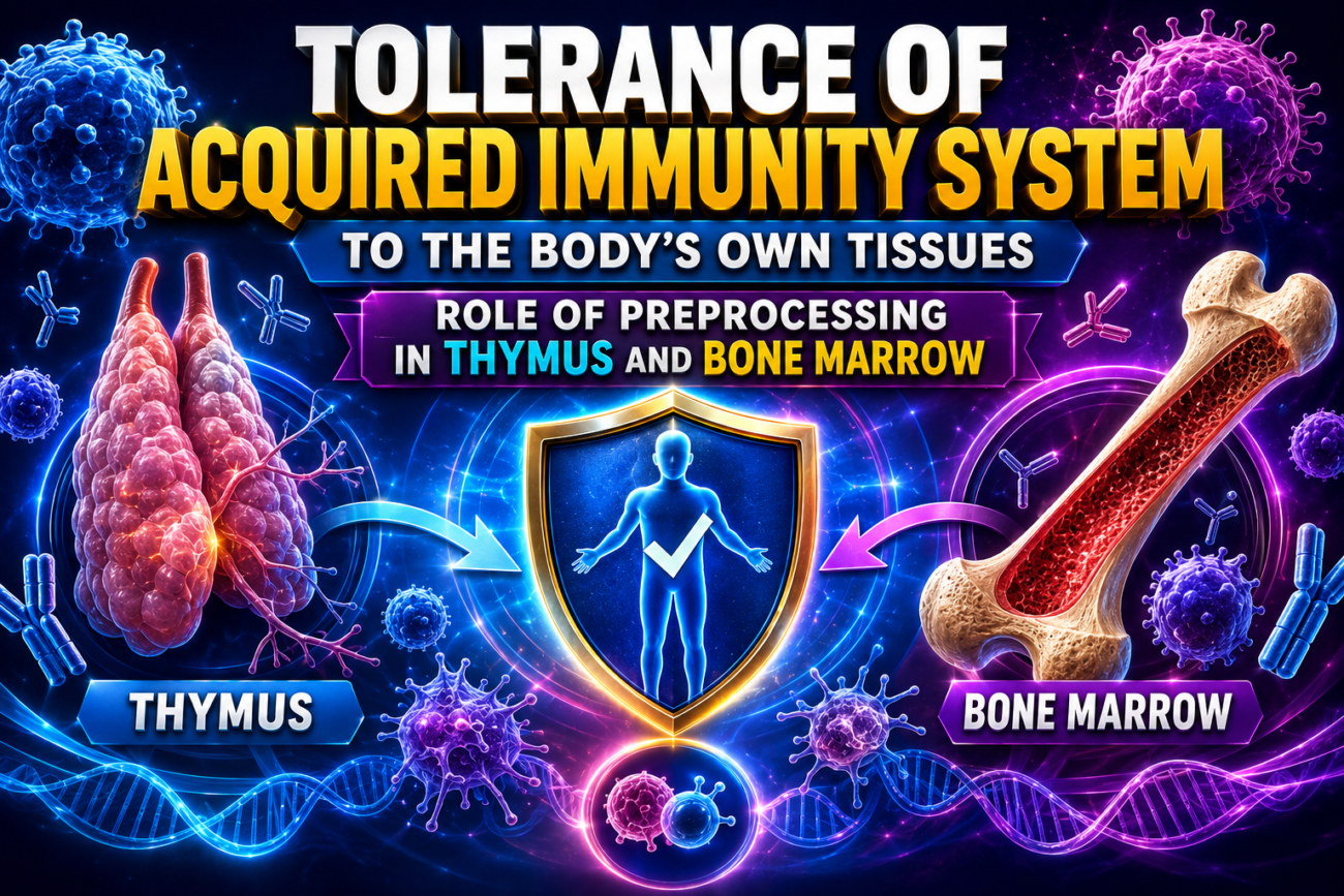
Tolerance of Acquired Immunity System to the Body’s Own Tissues—Role of Preprocessing in Thymus and Bone Marrow
- The acquired immune system could destroy the body’s own tissues if it reacted against them.
- Normally, the immune system recognizes the body’s own tissues as different from bacteria and viruses.
- Therefore, the immune system produces very few antibodies or activated T cells against the body’s own antigens.
Most Tolerance Results From Clone Selection During Preprocessing and Maturation
- Most immune tolerance develops during the preprocessing and maturation of:
- T lymphocytes in the thymus
- B lymphocytes in the bone marrow
- If a strong antigen is injected into a fetus while lymphocytes are being preprocessed, lymphocyte clones specific for that antigen fail to develop.
- Experiments show that immature T lymphocytes in the thymus respond to a strong antigen by:
- Becoming lymphoblastic
- Proliferating
- Combining with the stimulating antigen
- These lymphocytes are then destroyed by thymic epithelial cells.
- They are eliminated before migrating to the body’s lymphoid tissues.
- This process is called negative selection.
- Negative selection removes developing T lymphocytes and B lymphocytes that react against the body’s own tissues.
- These self-reactive lymphocytes are called autoreactive lymphocytes.
- During preprocessing in the thymus and bone marrow, most autoreactive lymphocyte clones are self-destroyed.
- This occurs because they are continuously exposed to the body’s own antigens.
- The thymus also produces regulatory T cells (Tregs).
- Tregs suppress autoreactive T lymphocytes that escape negative selection.
- Therefore, the thymus plays an essential role in developing immune tolerance.
Key Concept
Immune tolerance prevents the acquired immune system from attacking the body’s own tissues. During lymphocyte development, autoreactive T and B lymphocytes are eliminated through negative selection in the thymus and bone marrow. In addition, regulatory T cells generated in the thymus suppress any self-reactive T cells that escape deletion, making the thymus essential for maintaining self-tolerance and preventing autoimmunity.
Failure of the Tolerance Mechanism Causes Autoimmune Diseases
- Sometimes the body loses immune tolerance to its own tissues.
- This loss of tolerance becomes more common with increasing age.
- It often occurs after damage or destruction of the body’s own tissues.
- Tissue damage releases large amounts of self-antigens into the circulation.
- These self-antigens may stimulate the acquired immune system.
- As a result, the body produces:
- Activated T cells
- Antibodies
against its own tissues.
- More than 100 autoimmune diseases have been identified.
- Examples include:
- 1. Rheumatic Fever
- The immune system attacks the joints and heart, especially the heart valves.
- This occurs after exposure to a specific streptococcal toxin.
- The toxin contains an epitope that is similar to the body’s own self-antigens.
- 2. Glomerulonephritis (One Type)
- The immune system attacks the basement membrane of the glomeruli.
- 3. Myasthenia Gravis
- The immune system attacks acetylcholine receptor proteins at the neuromuscular junction.
- This results in paralysis.
- 4. Multiple Sclerosis (MS)
- The immune system attacks the myelin sheath covering nerve fibers.
- This disrupts communication within the nervous system.
- 5. Systemic Lupus Erythematosus (SLE)
- The immune system attacks many different body tissues simultaneously.
- Severe SLE can cause extensive tissue damage and may be fatal.
Figure Number
- Figure 35.9: Shows:
- Cytotoxic T cells (killer cells) binding to a target cell through specific antigen receptors.
- Release of cytotoxic and digestive enzymes.
- Destruction of the attacked cell.
Key Concept
Autoimmune diseases develop when immune tolerance fails and the immune system attacks the body’s own tissues. Tissue injury may release self-antigens that activate T cells and antibodies against self. Examples include rheumatic fever, glomerulonephritis, myasthenia gravis, multiple sclerosis (MS), and systemic lupus erythematosus (SLE).
Immunization by Injection of Antigens
- Immunization has been used for many years to produce acquired immunity against specific diseases.
- One method of immunization is the injection of dead organisms.
- These dead organisms cannot cause disease.
- However, they still contain antigens that stimulate the immune system.
- This type of immunization is used to protect against:
- Typhoid fever
- Whooping cough
- Diphtheria
- Many other bacterial diseases
- Immunity can also be produced by injecting toxins treated with chemicals.
- Chemical treatment destroys the toxic effect of the toxin.
- The antigenic properties of the toxin remain intact.
- This method is used to protect against:
- Tetanus
- Botulism
- Other similar toxin-mediated diseases
- Another method is immunization with live attenuated organisms.
- Attenuated organisms are weakened organisms that do not cause disease.
- They are produced by:
- Growing them in special culture media
- Passing them through a series of animals until they mutate
- Although weakened, they still contain the specific antigens needed to stimulate immunity.
- This method is used to protect against:
- Smallpox
- Yellow fever
- Poliomyelitis
- Measles
- Many other viral diseases
mRNA-Based Vaccines
- mRNA vaccines contain synthetic messenger RNA (mRNA) molecules.
- The synthetic mRNA instructs cells to produce a specific antigen.
- The produced antigen stimulates an immune response against a specific pathogen.
- Because mRNA is large and negatively charged, it cannot pass directly through cell membranes.
- Lipid-based nanoparticles are used to deliver the mRNA into cells.
- These nanoparticles fuse with the cell membrane.
- They form endosomes that enter the cytoplasm.
- The mRNA then escapes from the endosome into the cytoplasm.
- Inside antigen-presenting cells, ribosomes translate the mRNA into antigen protein.
- The antigen activates the immune system by:
- Stimulating B lymphocytes to produce antibodies.
- Activating phagocytic macrophages.
- Activating cytotoxic T cells.
- Activating T-helper cells.
- During the COVID-19 pandemic in 2020, mRNA vaccines were rapidly developed against Severe Acute Respiratory Syndrome Coronavirus-2 (SARS-CoV-2).
- These mRNA vaccines have been administered worldwide.
- mRNA vaccine technology is also being studied for the prevention and treatment of other diseases.
Figure Number
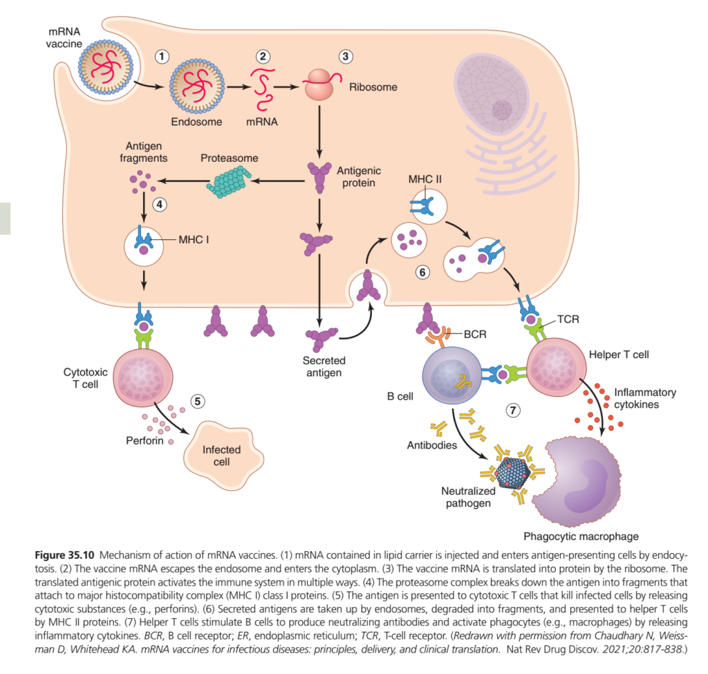
- Figure 35.10: Shows:
- Delivery of mRNA into cells by lipid-based nanoparticles
- Formation of endosomes
- Release of mRNA into the cytoplasm
- Production of antigen proteins by ribosomes
- Activation of:
- B lymphocytes
- Macrophages
- Cytotoxic T cells
- T-helper cells
Key Concept
Immunization produces acquired immunity by exposing the body to antigens through dead organisms, chemically inactivated toxins (toxoids), or live attenuated organisms. mRNA vaccines use synthetic mRNA delivered by lipid nanoparticles to produce antigen proteins inside antigen-presenting cells, stimulating both humoral and cell-mediated immune responses.
Passive Immunity
- The immunity discussed previously is active immunity.
- In active immunity, the person’s own body produces:
- Antibodies
- Activated T cells
in response to a foreign antigen.
- Passive immunity provides temporary protection.
- In passive immunity, no antigen is injected into the person.
- Instead, the person receives:
- Antibodies
- Activated T cells
- Or both
- These antibodies or activated T cells are obtained from:
- The blood of another person who has been actively immunized
- Another animal that has been actively immunized against the same antigen
- The transferred antibodies remain in the recipient’s body for about 2 to 3 weeks.
- During this period, the recipient is protected against the disease.
- The transfer of antibodies or activated T lymphocytes from another individual to provide protection is called passive immunity.
Key Concept
Passive immunity provides immediate but temporary protection by transferring preformed antibodies, activated T cells, or both from an actively immunized person or animal. Unlike active immunity, the recipient does not produce their own immune response, and protection usually lasts only 2–3 weeks.
Guyton Physiology Figure 35.10 Explained in Detail
Mechanism of Action of mRNA Vaccines (How mRNA Vaccines Produce Immunity)
This figure explains how mRNA vaccines (such as the Pfizer-BioNTech and Moderna COVID-19 vaccines) stimulate the immune system without using a live virus or even a whole virus particle.
Instead of injecting an antigen directly, an mRNA vaccine provides the genetic instructions (mRNA) that allow our own cells to temporarily produce a harmless viral protein (antigen). This protein then activates both humoral immunity (B cells and antibodies) and cell-mediated immunity (T cells).
This figure is extremely important because it integrates almost all the concepts you have learned in previous figures:
- Antigen presentation
- MHC Class I and II
- Cytotoxic T cells
- Helper T cells
- B cells
- Plasma cells
- Antibody production
- Macrophage activation
Overall Concept
Imagine a factory.
Instead of sending a finished product,
the company sends only the instruction manual.
The workers inside the factory read the manual,
manufacture the product,
display it,
and the security team learns how to recognize it.
Exactly the same thing happens with an mRNA vaccine.
- mRNA = Instruction manual
- Cell = Factory
- Ribosome = Machine
- Protein = Vaccine antigen
- Immune system = Security team
The vaccine does not contain the virus and cannot cause COVID-19.
It only teaches the immune system how to recognize the virus.
First Look at the Figure
The figure is divided into seven numbered steps.
1. mRNA vaccine enters cell
          ↓
2. mRNA escapes into cytoplasm
          ↓
3. Ribosomes produce antigenic protein
          ↓
4. MHC I presents antigen → Cytotoxic T cells
          ↓
5. Cytotoxic T cells destroy infected cells
          ↓
6. MHC II presents antigen → Helper T cells
          ↓
7. Helper T cells activate B cells
          ↓
Antibodies + Memory cells + Macrophage activation
Notice that one vaccine activates both arms of adaptive immunity.
Step 1
Entry of the mRNA Vaccine
Look at the left side of the figure.
The vaccine is enclosed inside a
Lipid Nanoparticle (LNP)
The lipid coat protects the fragile mRNA.
Without this lipid layer,
the mRNA would be rapidly destroyed by enzymes called RNases.ow Does the Vaccine Enter the Cell?
The cell membrane surrounds the lipid nanoparticle.
The cell engulfs it by
Endocytosis
forming an
Endosome.
Easy Analogy
Imagine receiving a parcel.
The delivery box is taken inside your house.
The cardboard box represents the
Endosome.
Step 2
mRNA Escapes from the Endosome
Inside the endosome,
the lipid nanoparticle breaks down.
The
mRNA
escapes into the
Cytoplasm.
Important Point
Notice something very important.
The mRNA
never enters the nucleus.
Therefore,
it
- does not mix with DNA
- does not alter human genes
- is eventually degraded naturally
Easy Analogy
The instruction manual stays in the factory floor.
It never enters the company’s central archive (the nucleus).Step 3
Translation by Ribosomes
The figure shows
Ribosome
Function
The ribosome reads the mRNA.
↓
Produces
the
Antigenic Protein
For COVID-19 vaccines,
this protein is the
Spike (S) protein.
Easy Analogy
The ribosome is like a 3D printer.
The mRNA is the blueprint.
The printed object is the viral protein.
Important Concept
The vaccine does not produce the virus.
It produces only
one harmless viral protein.
What Happens to the Antigenic Protein?
The newly synthesized protein has two major pathways.
One pathway activates
Cytotoxic T cells.
The other activates
Helper T cells and B cells.
Step 4
MHC Class I Presentation
Some antigenic protein remains inside the cell.
The
Proteasome
cuts it into
small peptide fragments.
Proteasome
Think of it as
a molecular shredder.
It cuts proteins into small peptides.
These peptides bind to
MHC Class I
inside the cell.
The peptide–MHC I complex is transported to the cell surface.
Who Recognizes MHC I?
The answer is
Cytotoxic T Cells (CD8⁺)
Easy Analogy
Imagine hanging a “Most Wanted” poster outside a building.
The poster alerts police officers.
MHC I is that poster.
Step 5
Cytotoxic T Cells Kill Infected Cells
The cytotoxic T cell recognizes the
Peptide–MHC I complex.
↓
It becomes activated.
↓
Releases
Perforin
and
Granzymes
↓
The infected cell undergoes
Apoptosis.
Why Is This Important?
If the real virus later infects body cells,
memory cytotoxic T cells will rapidly destroy those infected cells before the virus spreads extensively.
Clinical Example
During SARS-CoV-2 infection,
vaccinated individuals develop memory CD8⁺ T cells that help eliminate infected cells, reducing severe disease.
Step 6
MHC Class II Presentation
Some antigenic protein is
released outside the cell.
Nearby
Antigen-Presenting Cells (APCs)
such as dendritic cells and macrophages
engulf the protein.
↓
They digest it.
↓
Peptides bind to
MHC Class II.
Who Recognizes MHC II?
The answer is
Helper T Cells (CD4⁺)
Easy Analogy
MHC II is like a teacher showing a picture to the class.
The helper T cell studies the picture and plans the immune response.
Step 7
Activation of Helper T Cells
The helper T cell recognizes
Peptide + MHC II.
↓
Becomes activated.
↓
Releases
Cytokines
Examples include:
- IL-2
- IL-4
- IL-5
- IL-21
- IFN-γ
These cytokines coordinate the immune response.
Activation of B Cells
The figure shows a
B Cell
with its
B-Cell Receptor (BCR)
The BCR binds the antigen.
Helper T cells provide cytokines and co-stimulatory signals.
↓
B cells undergo
Clonal Expansion.
↓
Differentiate into
Plasma Cells.
Plasma Cells Produce Antibodies
Plasma cells produce
neutralizing antibodies.
These antibodies bind the viral spike protein.
↓
The virus can no longer attach to host cells.
↓
The pathogen becomes
Neutralized.
Easy Analogy
Imagine covering every keyhole with glue.
The key can no longer enter.
Neutralizing antibodies prevent the virus from entering cells.
Activation of Macrophages
The helper T cells also release
Inflammatory Cytokines
These activate
Macrophages.
Activated macrophages:
- engulf pathogens
- remove dead cells
- produce additional cytokines
- enhance inflammation
Formation of Memory Cells
Although not specifically labeled in the figure,
the immune response also generates:
- Memory B cells
- Memory Helper T cells
- Memory Cytotoxic T cells
These remain in the body for years.
Upon future exposure,
they respond rapidly.
This is the basis of
long-term vaccine protection.
Why Are mRNA Vaccines Effective?
One mRNA vaccine stimulates:
Cell-Mediated Immunity
✔ Cytotoxic T cells
✔ Memory T cells
Humoral Immunity
✔ Helper T cells
✔ B cells
✔ Plasma cells
✔ Neutralizing antibodies
✔ Memory B cells
Thus,
both major branches of adaptive immunity are activated.s the Vaccine Change DNA?
A common misconception is that mRNA vaccines alter human DNA.
They do not.
Reasons:
- mRNA remains in the cytoplasm.
- It does not enter the nucleus.
- Human ribosomes translate it into protein.
- The mRNA is naturally degraded within a short time.
Clinical Correlations
1. COVID-19 Vaccination
The Pfizer-BioNTech and Moderna vaccines deliver mRNA encoding the SARS-CoV-2 spike protein.
The immune system develops neutralizing antibodies and T-cell responses that reduce the risk of severe COVID-19.2. Booster Doses
Booster vaccines reactivate memory B cells and T cells,
leading to a rapid, high-level secondary immune response.
3. Future Applications
mRNA technology is being investigated for:
- Influenza vaccines
- RSV vaccines
- Personalized cancer vaccines
- Vaccines against emerging infectious diseases
Relationship with Previous Figures
| Figure | Main Concept |
|---|---|
| 35.1 | Development of T and B lymphocytes |
| 35.2 | Clonal selection of B cells |
| 35.3 | Primary and secondary immune responses |
| 35.4 | Structure of antibodies |
| 35.5 | Agglutination by antibodies |
| 35.6 | Complement activation |
| 35.8 | Helper T cells regulate immunity |
| 35.9 | Cytotoxic T cells destroy infected cells |
| 35.10 | mRNA vaccines activate both humoral and cell-mediated immunity |
Notice how Figure 35.10 combines almost every concept from the previous figures into one integrated immune response.
High-Yield MBBS Points
- mRNA vaccines deliver messenger RNA, not live virus.
- The mRNA is protected by a lipid nanoparticle and enters cells by endocytosis.
- The mRNA escapes into the cytoplasm, where ribosomes translate it into an antigenic protein.
- The antigen is processed and presented on MHC Class I, activating CD8⁺ cytotoxic T cells.
- Secreted antigen is taken up by APCs and presented on MHC Class II, activating CD4⁺ helper T cells.
- Helper T cells stimulate B-cell proliferation, plasma-cell formation, and antibody production.
- Neutralizing antibodies prevent the pathogen from entering host cells.
- Memory B cells and memory T cells provide long-term protection.
- mRNA does not enter the nucleus and does not alter human DNA.
Complete Flow Chart
mRNA Vaccine Injection
          ↓
Lipid Nanoparticle Enters Cell
          ↓
Endocytosis
          ↓
mRNA Released into Cytoplasm
          ↓
Ribosome Produces Antigenic Protein
          ↓
 ┌─────────────────────────────┐
 │                             │
 ▼                             ▼
Proteasome                 Secreted Antigen
 │                             │
 ▼                             ▼
MHC Class I               APC Engulfs Antigen
 │                             │
 ▼                             ▼
CD8⁺ Cytotoxic T Cell     MHC Class II
 │                             │
 ▼                             ▼
Kills Infected Cells      CD4⁺ Helper T Cell
                                │
               ┌────────────────┴────────────────┐
               ▼                                 ▼
           B-Cell Activation              Macrophage Activation
               │
               ▼
          Plasma Cells
               │
               ▼
     Neutralizing Antibodies
               │
               ▼
Memory B Cells + Memory T Cells
               │
               ▼
Long-Term Protective Immunity
Key Concept
Figure 35.10 demonstrates how mRNA vaccines generate protective immunity without using a live virus. After entering host cells, the vaccine mRNA is translated into a harmless antigenic protein. This protein is presented through both MHC Class I and MHC Class II pathways, activating CD8⁺ cytotoxic T cells and CD4⁺ helper T cells. Helper T cells stimulate B cells to produce neutralizing antibodies and activate macrophages through cytokine release. At the same time, memory B cells and memory T cells are generated, providing rapid and long-lasting protection against future infection.
Tolerance of the Acquired Immune System to the Body’s Own Tissues, Autoimmunity, Immunization, and Passive Immunity ( Summary)
The acquired immune system protects the body from harmful microorganisms such as bacteria and viruses. However, if it were unable to distinguish between foreign substances and the body’s own tissues, it would attack normal cells and cause serious damage. To prevent this, the immune system develops immune tolerance, which is the ability to recognize the body’s own tissues as “self” and avoid attacking them. As a result, healthy tissues are normally protected from immune destruction.
Most immune tolerance develops while lymphocytes are still immature. T lymphocytes mature in the thymus, whereas B lymphocytes mature in the bone marrow. During this developmental stage, these cells undergo a strict selection process. If an immature T or B lymphocyte recognizes and reacts strongly against the body’s own antigens (self-antigens), it is eliminated before it can enter the circulation. This process is known as negative selection. In this way, potentially harmful self-reactive lymphocytes are destroyed, allowing only safe lymphocytes to mature and participate in immune defense.
The thymus also produces Regulatory T cells (Tregs), which play an important role in maintaining immune tolerance. These specialized cells suppress any self-reactive T lymphocytes that may escape negative selection, preventing them from attacking the body’s own tissues. Therefore, the thymus is essential for maintaining self-tolerance and preventing autoimmune diseases.
Sometimes the immune tolerance mechanism fails, causing the immune system to mistakenly attack normal body tissues. This condition is known as autoimmunity, and the diseases that result are called autoimmune diseases. Autoimmunity may occur when damaged tissues release self-antigens that are mistakenly recognized as foreign. The risk of developing autoimmune diseases generally increases with age.
Several important autoimmune diseases illustrate this abnormal immune response. In rheumatic fever, antibodies produced after a streptococcal infection mistakenly attack the heart valves and joints because the bacterial antigens resemble the body’s own tissues. In glomerulonephritis, the immune system attacks the basement membrane of the kidney glomeruli, leading to kidney damage. In myasthenia gravis, antibodies attack acetylcholine receptors at the neuromuscular junction, resulting in muscle weakness and paralysis. In multiple sclerosis (MS), the immune system destroys the myelin sheath surrounding nerve fibers, impairing nerve conduction. In systemic lupus erythematosus (SLE), the immune system attacks multiple organs and tissues simultaneously, causing widespread inflammation and potentially life-threatening damage.
To protect people from infectious diseases, immunization is used to stimulate acquired immunity before natural infection occurs. Immunization introduces antigens into the body, allowing the immune system to produce antibodies, activate T cells, and develop memory cells for long-term protection. Vaccines may contain killed (dead) microorganisms, toxoids (chemically inactivated bacterial toxins), or live attenuated organisms that have been weakened so they cannot cause disease but still stimulate immunity. These vaccines provide effective protection against many bacterial and viral diseases.
A modern approach to immunization is the mRNA vaccine. Instead of containing the antigen itself, these vaccines contain synthetic messenger RNA (mRNA) enclosed within lipid nanoparticles. After entering body cells, the mRNA is translated by ribosomes into antigen proteins. These proteins are recognized by the immune system, stimulating antibody production by B lymphocytes, activation of helper and cytotoxic T cells, and increased activity of macrophages. The rapid development of mRNA vaccines against COVID-19 (SARS-CoV-2) demonstrated the effectiveness of this technology, which is now being investigated for the prevention and treatment of many other diseases.
The immunity produced after vaccination is called active immunity because the person’s own immune system generates antibodies, activated T cells, and memory cells. Although active immunity takes time to develop, it usually provides long-lasting protection.
In contrast, passive immunity provides immediate but temporary protection. Instead of stimulating the person’s immune system, ready-made antibodies or activated T cells obtained from another individual or an immunized animal are transferred directly into the recipient. These antibodies provide instant defense against infection but remain in the body for only 2–3 weeks, and no memory cells are formed. Consequently, passive immunity offers rapid yet short-term protection and is often used when immediate immunity is required.
In summary, immune tolerance protects the body from attacking its own tissues through negative selection of self-reactive lymphocytes and the action of regulatory T cells. Failure of this tolerance leads to autoimmune diseases. Immunization safely stimulates active immunity and long-lasting protection, while passive immunity provides immediate but temporary protection through the transfer of ready-made antibodies or activated T cells. These mechanisms together form the foundation of the body’s acquired immune defense system.
MADE BY SELF LEARNING CEO AND FOUNDER DR SHEEN
