


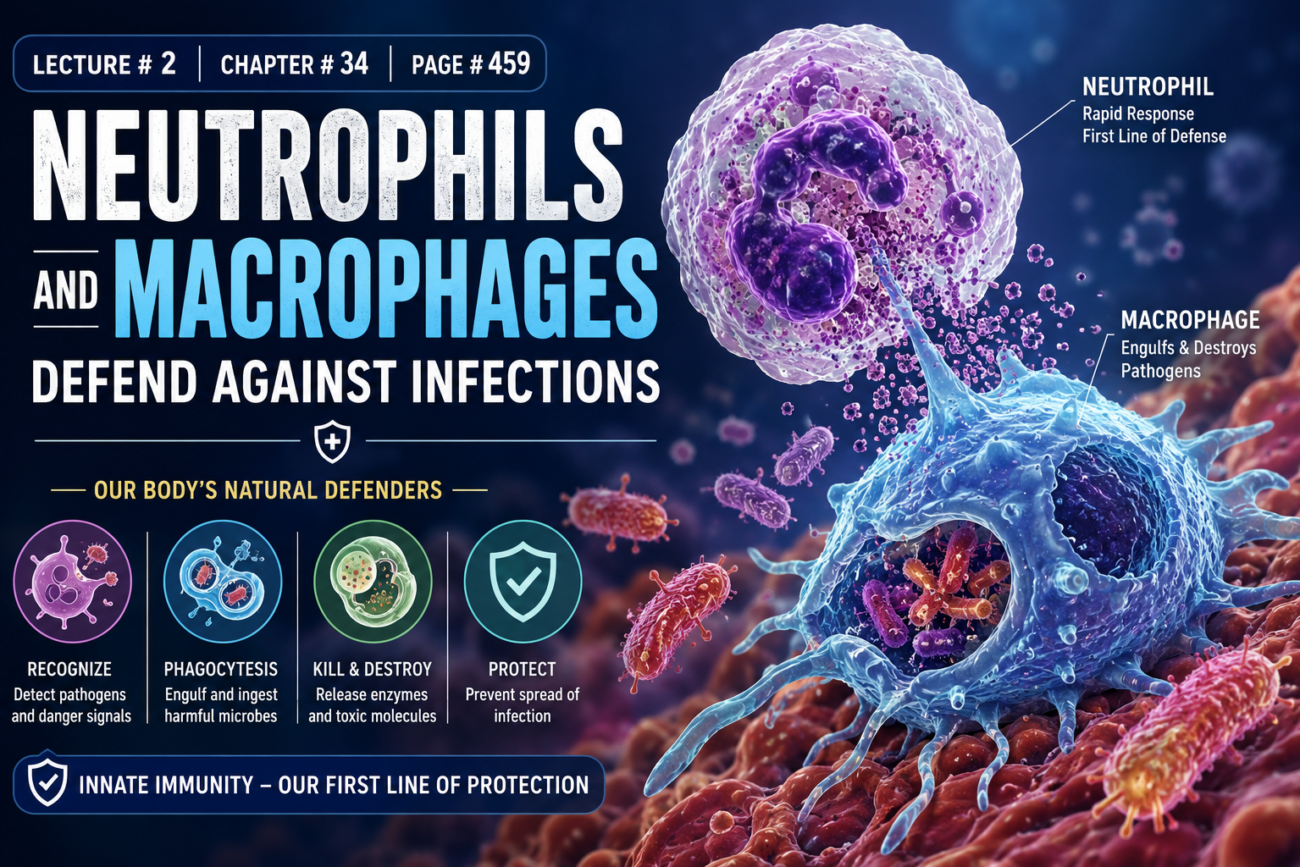
- Neutrophils and tissue macrophages are the main cells that attack and destroy:
- Bacteria
- Viruses
- Other harmful agents
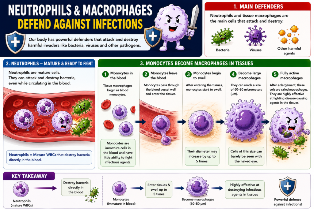
- Neutrophils are mature cells.
- They can attack and destroy bacteria, even while circulating in the blood.
- Tissue macrophages begin as blood monocytes.
- Monocytes are immature cells while they are in the blood.
- At this stage, monocytes have little ability to fight infectious agents.
- After monocytes enter the tissues:
- They begin to swell.
- Their diameter may increase by up to 5 times.
- They can reach a size of 60–80 micrometers (µm).
- Cells of this size can barely be seen with the naked eye.
- After enlargement, these cells are called macrophages.
- Macrophages are highly effective at fighting disease-causing agents in the tissues.
Key Concept
Neutrophils are mature WBCs that destroy bacteria directly in the blood. Monocytes enter the tissues, enlarge up to 5 times (60–80 µm), become macrophages, and become highly effective cells for destroying infectious agents in the tissues.
White Blood Cells Enter Tissue Spaces and Move by Extravastion (Diapedesis) and Ameboid Motion
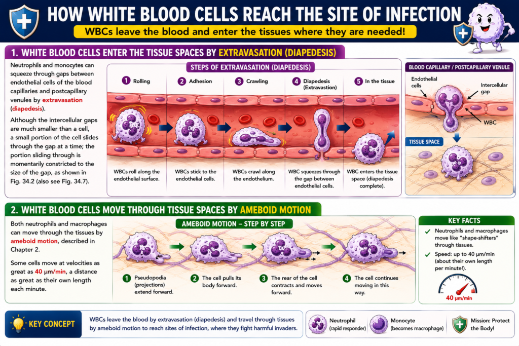
- Neutrophils and monocytes can leave the blood vessels by extravasation (diapedesis).
- During extravasation (diapedesis), they squeeze through the gaps between endothelial cells of:
- Blood capillaries
- Postcapillary venules
- The gaps between endothelial cells are much smaller than the white blood cell.
- A small part of the cell passes through the gap at a time.
- The part passing through is temporarily compressed to fit the size of the gap.
- This process is shown in Figure 34.2.
- It is also shown in Figure 34.7.
- After entering the tissues, neutrophils and macrophages move by ameboid motion.
- Ameboid motion was described in Chapter 2.
- Some white blood cells can move at a speed of 40 µm/min.
- This distance is approximately equal to their own body length every minute.
Figure
- Figure 34.2 – White blood cells passing through endothelial gaps by extravasation (diapedesis).
- Figure 34.7 – Also shows extravasation (diapedesis).
Equations
- No mathematical equation is present in the provided text.
Key Concept
Neutrophils and monocytes leave the bloodstream by extravasation (diapedesis), squeezing through tiny endothelial gaps. Once inside the tissues, neutrophils and macrophages move by ameboid motion, reaching speeds of up to 40 µm/min.
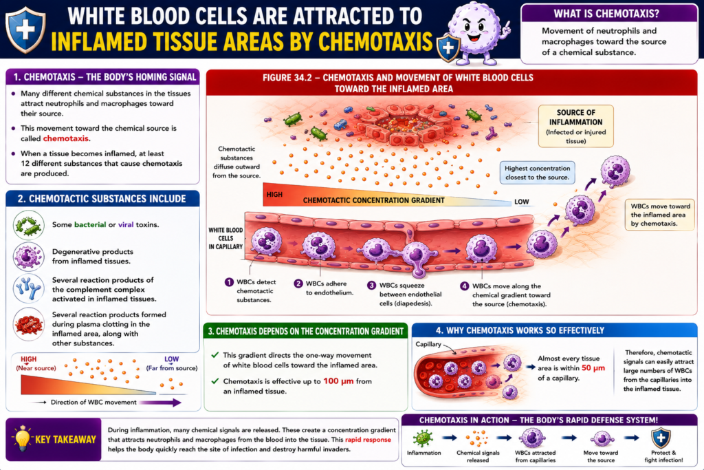
White Blood Cells Are Attracted to Inflamed Tissue Areas by Chemotaxis
- Many different chemical substances in the tissues attract neutrophils and macrophages toward their source.
- This movement toward the chemical source is called chemotaxis.
- Chemotaxis is shown in Figure 34.2.
- When a tissue becomes inflamed, at least 12 different substances that cause chemotaxis are produced.
- These chemotactic substances include:
- Some bacterial or viral toxins.
- Degenerative products from inflamed tissues.
- Several reaction products of the complement complex activated in inflamed tissues.
- Several reaction products formed during plasma clotting in the inflamed area, along with other substances.
- Chemotaxis depends on the concentration gradient of the chemotactic substance.
- The highest concentration is closest to the source.
- This concentration gradient directs the one-way movement of white blood cells toward the inflamed area.
- Chemotaxis is effective up to 100 µm from an inflamed tissue.
- Almost every tissue area is within 50 µm of a capillary.
- Therefore, chemotactic signals can easily attract large numbers of WBCs from the capillaries into the inflamed tissue.
Figure
- Figure 34.2 – Chemotaxis and movement of white blood cells toward the inflamed area.
Equations
- No mathematical equation is present in the provided text.
Key Concept
Chemotaxis is the movement of neutrophils and macrophages toward an inflamed area in response to a chemical concentration gradient. Chemotactic substances produced during inflammation attract WBCs from nearby capillaries, allowing them to quickly reach and defend the infected tissue.
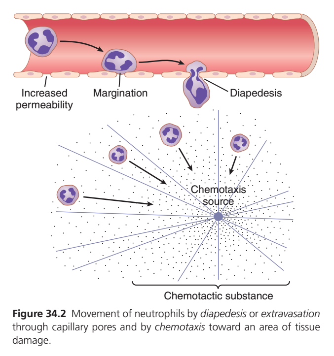
Movement of Neutrophils to an Infection Site (Guyton Figure 34.2) – Easiest Concept
One Main Idea
When tissue is injured or infected, neutrophils quickly leave the bloodstream and move to the damaged area to destroy bacteria.
This process occurs in 4 simple steps:
Inflammation
      ↓
Margination
      ↓
Diapedesis
      ↓
Chemotaxis
      ↓
Neutrophils Reach Infection
      ↓
Destroy Bacteria (Phagocytosis)
Step 1. Increased Permeability
What happens?
During inflammation, blood vessels become:
- Wider (vasodilation)
- More permeable (leaky)
This allows white blood cells to leave the blood more easily.
Easy Concept
Think of the blood vessel as a road.
Normally, the road has closed gates.
During inflammation, the gates open, allowing neutrophils to exit.
Step 2. Margination
What is Margination?
Normally, neutrophils flow in the center of the blood vessel.
During inflammation, blood flow slows down.
As a result, neutrophils move and stick to the inner wall of the blood vessel.
This is called:
Margination
Easy Definition
Margination = Neutrophils move from the center of the blood vessel to its wall.
Memory Trick
Margin = Edge
➡️ Margination = Moving to the edge (wall).
Step 3. Diapedesis (Extravasation)
What is Diapedesis?
After sticking to the vessel wall, neutrophils squeeze between endothelial cells and leave the blood vessel.
This movement is called:
Diapedesis (also called Extravasation).
Easy Definition
Diapedesis = WBCs squeeze through capillary walls into the tissues.
Memory Trick
D = Door
Diapedesis means the white blood cell passes through the “door” of the blood vessel.
Step 4. Chemotaxis
What is Chemotaxis?
Damaged tissue and bacteria release special chemicals called chemotactic substances.
These chemicals create a chemical gradient.
Neutrophils detect this gradient and move toward the highest concentration, where the infection is located.
This directed movement is called:
Chemotaxis
Easy Definition
Chemotaxis = Movement of neutrophils toward the source of infection by following chemical signals.
Memory Trick
Chemo = Chemical
Taxis = Movement
➡️ Chemotaxis = Movement toward chemicals.
What Happens After Chemotaxis?
Once neutrophils reach the infected tissue, they:
- Surround bacteria
- Engulf bacteria (Phagocytosis)
- Destroy bacteria using enzymes and reactive oxygen species
This helps stop the infection.
Complete Sequence
Injury or Infection
        ↓
Inflammation
        ↓
Blood vessels become leaky
(Increased permeability)
        ↓
Neutrophils move to vessel wall
(Margination)
        ↓
Neutrophils squeeze out of vessel
(Diapedesis)
        ↓
Neutrophils follow chemical signals
(Chemotaxis)
        ↓
Reach infection site
        ↓
Phagocytosis of bacteria
Easy Analogy
Imagine there is a fire in a building.
- 🚒 Blood vessel = Road
- 👨🚒 Neutrophils = Firefighters
- 🚪 Diapedesis = Firefighters leaving the road through a gate
- 📍 Chemotactic chemicals = Smoke guiding firefighters
- 🔥 Infection = Fire
The firefighters follow the smoke until they reach the fire and put it out.
Key Terms at a Glance
| Term | Easy Meaning |
|---|---|
| Increased Permeability | Blood vessels become leaky so WBCs can leave. |
| Margination | Neutrophils move to the edge (wall) of the blood vessel. |
| Diapedesis (Extravasation) | Neutrophils squeeze through the vessel wall into tissues. |
| Chemotaxis | Neutrophils follow chemical signals to the site of infection. |
| Phagocytosis | Neutrophils engulf and destroy bacteria. |
One-Line Revision
Infection → Increased permeability → Margination → Diapedesis → Chemotaxis → Phagocytosis
Exam Pearls (Guyton)
- Margination is the movement of neutrophils from the center of the bloodstream to the vessel wall.
- Diapedesis (extravasation) is the passage of neutrophils through capillary pores into tissues.
- Chemotaxis is the directed movement of neutrophils toward chemicals released by damaged tissues, bacteria, and inflammatory mediators.
- Increased vascular permeability allows white blood cells and plasma proteins to leave the circulation and enter inflamed tissue.
- Neutrophils are the first white blood cells to arrive at the site of acute inflammation and begin phagocytosis of microorganisms.
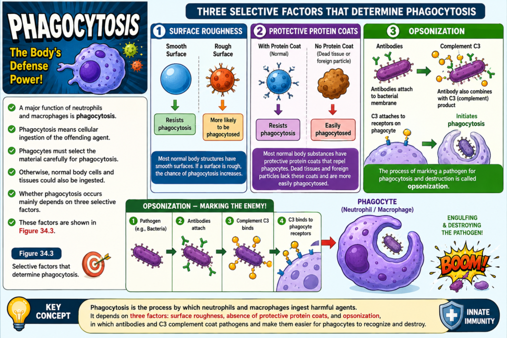
Phagocytosis
- A major function of neutrophils and macrophages is phagocytosis.
- Phagocytosis means cellular ingestion of the offending agent.
- Phagocytes must select the material carefully for phagocytosis.
- Otherwise, normal body cells and tissues could also be ingested.
- Whether phagocytosis occurs mainly depends on three selective factors.
- These factors are shown in Figure 34.3.
- First factor:
- Most normal body structures have smooth surfaces.
- Smooth surfaces resist phagocytosis.
- If a surface is rough, the chance of phagocytosis increases.
- Second factor:
- Most normal body substances have protective protein coats.
- These protein coats repel phagocytes.
- Most dead tissues and foreign particles do not have protective protein coats.
- Therefore, they are more easily phagocytosed.
- Third factor:
- The immune system produces antibodies against infectious agents such as bacteria.
- The antibodies attach to the bacterial membrane.
- This makes the bacteria more susceptible to phagocytosis.
- The antibody also combines with the C3 product of the complement cascade.
- C3 molecules attach to receptors on the phagocyte membrane.
- This attachment initiates phagocytosis.
- The process of marking a pathogen for phagocytosis and destruction is called opsonization.
Figure
- Figure 34.3 – Selective factors that determine phagocytosis.
Equations
- No mathematical equation is present in the provided text.
Key Concept
Phagocytosis is the process by which neutrophils and macrophages ingest harmful agents. It depends on three factors: surface roughness, absence of protective protein coats, and opsonization, in which antibodies and C3 complement coat pathogens and make them easier for phagocytes to recognize and destroy.
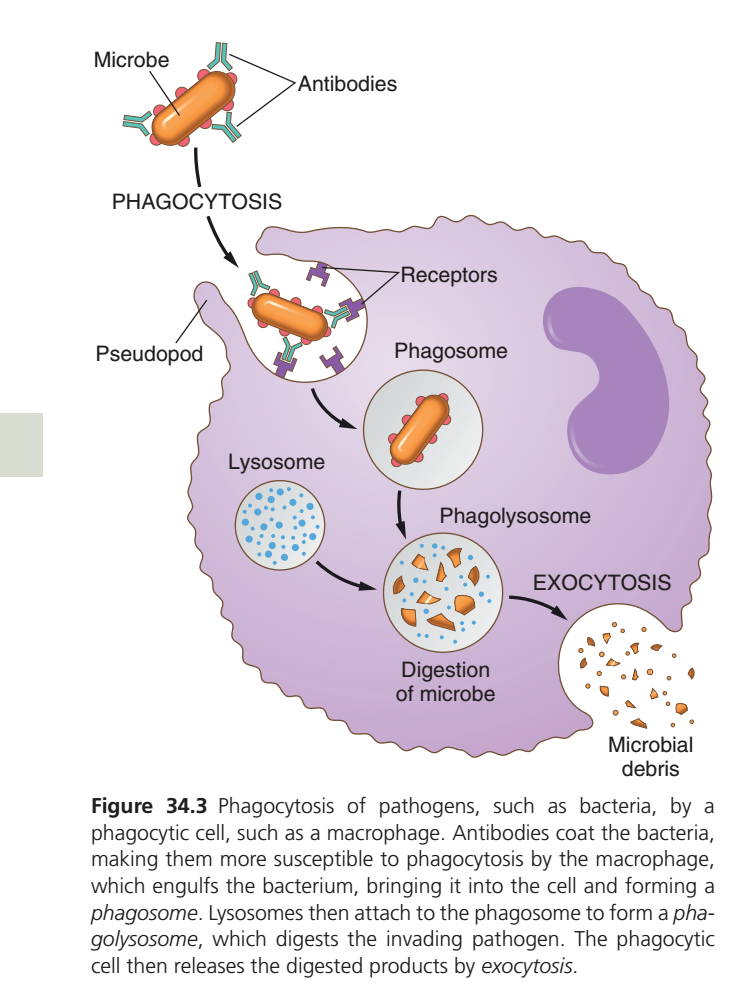
Mechanism of Phagocytosis (Guyton Figure 34.3) – Easiest Conceptual Summary
One Main Idea
Phagocytosis is the process in which a neutrophil or macrophage recognizes, engulfs, digests, and destroys a microbe.
Think of a phagocyte as the body’s security guard and garbage collector. It catches the invader, destroys it, and throws away the waste.
Complete Process in 6 Easy Steps
Microbe enters body
        ↓
1. Recognition & Attachment
        ↓
2. Engulfment (Pseudopods)
        ↓
3. Phagosome Formation
        ↓
4. Phagolysosome Formation
        ↓
5. Digestion of Microbe
        ↓
6. Exocytosis (Waste Removal)
Step 1. Recognition and Attachment
What happens?
The bacterium is first coated with antibodies (and often complement proteins such as C3).
This process is called:
Opsonization
The phagocyte has receptors on its surface that recognize these antibodies and complement proteins.
Once recognized, the phagocyte firmly attaches to the bacterium.
Easy Concept
Think of antibodies as “name tags” placed on bacteria.
The phagocyte reads the tag and knows exactly which cell to attack.Step 2. Engulfment (Pseudopods)
After attachment:
The phagocyte extends pseudopods (“false feet”) around the bacterium.
These pseudopods slowly surround the microbe until it is completely enclosed.
Easy Definition
Pseudopods = Temporary arm-like extensions used to catch and surround microbes.
Memory Trick
Pseudo = False
Pod = Foot
➡️ Pseudopod = False foot
Step 3. Formation of the Phagosome
Once the bacterium is fully enclosed:
It becomes trapped inside a membrane-bound sac called the:
Phagosome
Easy Definition
Phagosome = A bubble inside the phagocyte containing the swallowed microbe.
Memory Trick
Phagosome = Food bag
The bacterium has been swallowed but is not yet destroyed.
Step 4. Formation of the Phagolysosome
Inside the cell are many lysosomes.
Lysosomes contain powerful digestive enzymes.
The lysosome fuses with the phagosome.
This forms a new structure called the:
Phagolysosome
Easy Definition
Phagolysosome = Phagosome + Lysosome
This is the place where the microbe is destroyed.
Memory Trick
Lysosome = Lyses (breaks down) everything.
Step 5. Digestion of the Microbe
Inside the phagolysosome:
- Digestive enzymes
- Toxic oxygen radicals
- Other antimicrobial substances
break down and kill the bacterium.
The microbe is converted into tiny harmless fragments.
Easy Concept
The phagolysosome acts like a stomach inside the cell.ep 6. Exocytosis (Waste Removal)
After digestion:
Only waste material remains.
The phagocyte pushes this waste outside the cell.
This process is called:
Exocytosis
Easy Definition
Exocytosis = Removal of digested waste from the cell.
Memory Trick
Exo = Exit
➡️ Waste exits the cell.
Whole Process in One Flow
Bacteria enters body
          ↓
Antibodies coat bacteria
(Opsonization)
          ↓
Phagocyte receptors recognize bacteria
          ↓
Pseudopods surround bacterium
          ↓
Phagosome forms
          ↓
Lysosome joins phagosome
          ↓
Phagolysosome forms
          ↓
Enzymes digest bacterium
          ↓
Microbial debris remains
          ↓
Exocytosis removes debris
Easy Analogy
Imagine a garbage truck cleaning a city.
- 🦠 Bacteria = Garbage
- 🏷️ Antibodies = Garbage label
- 🚛 Macrophage/Neutrophil = Garbage truck
- 🤲 Pseudopods = Truck’s grabbing arms
- 📦 Phagosome = Garbage container
- 🧪 Lysosome = Garbage crusher
- 🗑️ Exocytosis = Throwing crushed garbage away
Important Terms
| Term | Easy Meaning |
|---|---|
| Antibodies | Mark microbes so phagocytes can recognize them. |
| Receptors | Surface proteins on phagocytes that bind antibodies and complement. |
| Pseudopods | Temporary “false feet” that surround and engulf microbes. |
| Phagosome | Membrane-bound vesicle containing the engulfed microbe. |
| Lysosome | Organelle containing digestive enzymes. |
| Phagolysosome | Fusion of a phagosome and lysosome where the microbe is destroyed. |
| Digestion | Enzymes break the microbe into small fragments. |
| Exocytosis | The cell releases the remaining waste outside. |
Memory Tricks
Phagosome
“Food Packet”
The swallowed bacterium is stored here.
Lysosome
“Lysis = Breakdown”
It contains digestive enzymes.
Phagolysosome
“Destruction Chamber”
This is where microbes are killed and digested.
Exocytosis
Exo = Exit
Waste leaves the cell.
High-Yield Exam Points (Guyton)
- Antibodies and complement proteins (especially C3) coat microbes and make them easier for phagocytes to recognize (opsonization).
- Phagocyte receptors bind these coated microbes and initiate engulfment.
- Pseudopods surround the pathogen and enclose it within a phagosome.
- Lysosomes fuse with the phagosome to form a phagolysosome.
- Inside the phagolysosome, digestive enzymes and antimicrobial substances destroy the microorganism.
- The remaining microbial debris is expelled from the cell by exocytosis.
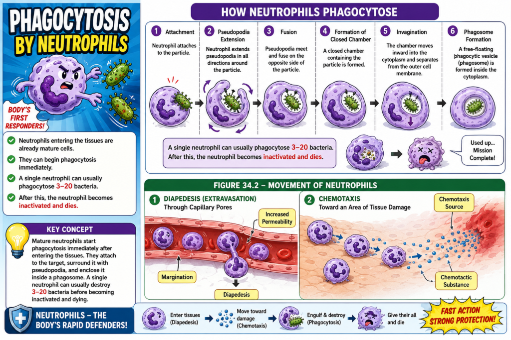
Phagocytosis by Neutrophils
- Neutrophils entering the tissues are already mature cells.
- They can begin phagocytosis immediately.
- When a neutrophil approaches a particle:
- It first attaches to the particle.
- The neutrophil then extends pseudopodia in all directions around the particle.
- The pseudopodia meet and fuse on the opposite side of the particle.
- This forms a closed chamber containing the particle.
- The chamber then moves inward (invaginates) into the cytoplasm.
- It separates from the outer cell membrane.
- This forms a free-floating phagocytic vesicle (phagosome) inside the cytoplasm.
- A single neutrophil can usually phagocytose 3–20 bacteria.
- After this, the neutrophil becomes inactivated and dies.
Figure
- Figure 34.2
- Movement of neutrophils by:
- Diapedesis (extravasation) through capillary pores.
- Chemotaxis toward an area of tissue damage.
- Labels shown in the figure:
- Chemotaxis source
- Chemotactic substance
- Increased permeability
- Margination
- Diapedesis
- Movement of neutrophils by:
Equations
- No mathematical equation is present in the provided text.
Key Concept
Mature neutrophils start phagocytosis immediately after entering the tissues. They attach to the target, surround it with pseudopodia, and enclose it inside a phagosome. A single neutrophil can usually destroy 3–20 bacteria before becoming inactivated and dying.
Phagocytosis by Macrophages
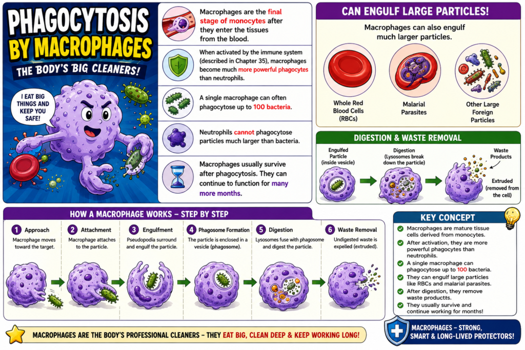
- Macrophages are the final stage of monocytes after they enter the tissues from the blood.
- When activated by the immune system (described in Chapter 35), macrophages become much more powerful phagocytes than neutrophils.
- A single macrophage can often phagocytose up to 100 bacteria.
- Macrophages can also engulf much larger particles.
- They can phagocytose:
- Whole red blood cells (RBCs)
- Occasionally, malarial parasites
- Neutrophils cannot phagocytose particles much larger than bacteria.
- After digesting the ingested particles, macrophages expel (extrude) the remaining waste products.
- Macrophages usually survive after phagocytosis.
- They can continue to function for many more months.
Figure
- No figure number is mentioned in the provided text.
Equations
- No mathematical equation is present in the provided text.
Key Concept
Macrophages are mature tissue cells derived from monocytes. After activation by the immune system, they become more powerful phagocytes than neutrophils, can ingest up to 100 bacteria, engulf large particles such as RBCs and malarial parasites, remove waste after digestion, and continue functioning for many months.
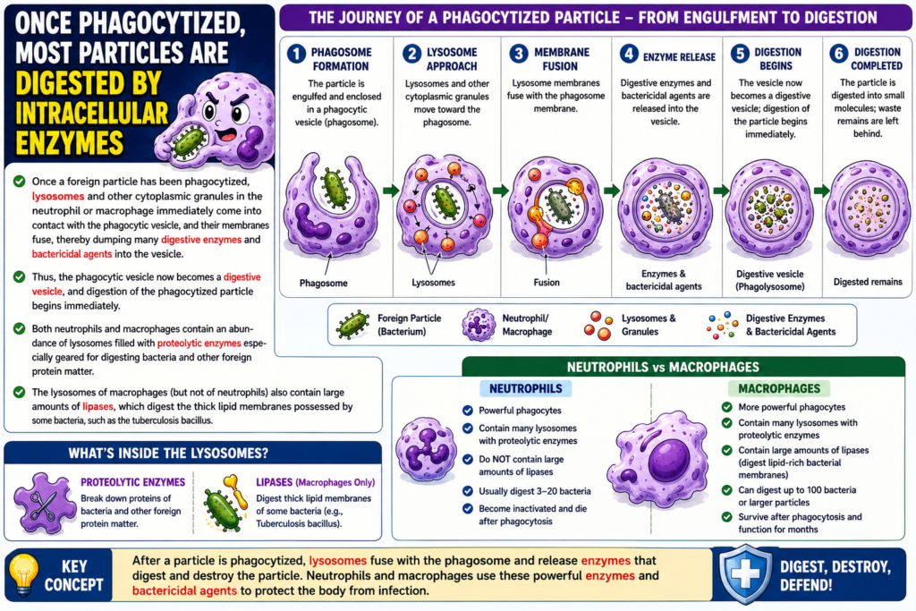
Once Phagocytized, Most Particles Are Digested by Intracellular Enzymes
- Once a foreign particle is phagocytized, lysosomes and other cytoplasmic granules in the neutrophil or macrophage immediately come into contact with the phagocytic vesicle.
- The membranes of the lysosomes and the phagocytic vesicle fuse together.
- This fusion releases digestive enzymes and bactericidal agents into the vesicle.
- The phagocytic vesicle is now converted into a digestive vesicle.
- Digestion of the phagocytized particle begins immediately.
- Both neutrophils and macrophages contain many lysosomes.
- These lysosomes are filled with proteolytic enzymes.
- Proteolytic enzymes digest:
- Bacteria
- Other foreign protein materials
- Macrophage lysosomes (but not neutrophil lysosomes) also contain large amounts of lipases.
- Lipases digest the thick lipid membranes of some bacteria.
- One example is the tuberculosis bacillus.
Figure
- No figure number is mentioned in the provided text.
Equations
- No mathematical equation is present in the provided text.
Key Concept
After phagocytosis, lysosomes fuse with the phagocytic vesicle, releasing digestive enzymes and bactericidal agents that immediately digest the ingested particle. Both neutrophils and macrophages use proteolytic enzymes, while macrophages also contain lipases that digest the lipid-rich membranes of bacteria such as the tuberculosis bacillus.
Neutrophils and Macrophages Can Kill Bacteria
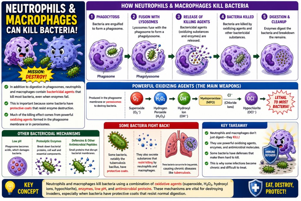
- In addition to digesting ingested bacteria inside phagosomes, neutrophils and macrophages contain bactericidal agents.
- These bactericidal agents can kill most bacteria, even when lysosomal enzymes fail to digest them.
- This ability is especially important because some bacteria have:
- Protective coats
- Other factors that prevent destruction by digestive enzymes
- Much of the bacterial killing is caused by powerful oxidizing agents.
- These oxidizing agents are formed by:
- Enzymes in the phagosome membrane
- A special organelle called the peroxisome
- The major oxidizing agents include:
- Superoxide (O₂⁻)
- Hydrogen peroxide (H₂O₂)
- Hydroxyl ions (OH⁻)
- These oxidizing agents are lethal to most bacteria, even in small amounts.
- One lysosomal enzyme, myeloperoxidase, catalyzes the reaction between:
- Hydrogen peroxide (H₂O₂)
- Chloride ions (Cl⁻)
- This reaction forms hypochlorite (OCl⁻).
- Hypochlorite is extremely bactericidal.
- Some bacteria, especially the tuberculosis bacillus, have protective coats that resist lysosomal digestion.
- These bacteria also secrete substances that partially resist the killing effects of neutrophils and macrophages.
- Such bacteria can cause chronic diseases, such as tuberculosis.
Figure
- No figure number is mentioned in the provided text.
Equations
- Reaction catalyzed by myeloperoxidase: H₂O₂ + Cl⁻ → Hypochlorite (OCl⁻)
- Important bactericidal oxidizing agents:
- Superoxide: O₂⁻
- Hydrogen peroxide: H₂O₂
- Hydroxyl ion: OH⁻
- Hypochlorite: OCl⁻
Key Concept
Neutrophils and macrophages kill bacteria not only by digesting them in phagosomes but also by using bactericidal oxidizing agents such as O₂⁻, H₂O₂, OH⁻, and hypochlorite (OCl⁻). Myeloperoxidase converts H₂O₂ and Cl⁻ into hypochlorite, a highly effective bactericidal substance. Some bacteria, such as the tuberculosis bacillus, resist these defense mechanisms and can cause chronic infections.
MADE BY SELF LEARNING CEO AND FOUNDER DR SHEEN
