


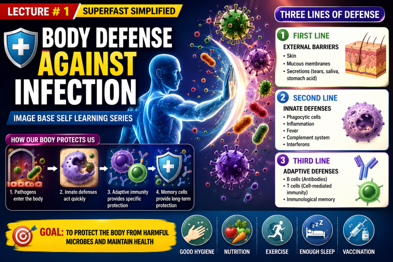
- Our body is constantly exposed to bacteria, viruses, fungi, and parasites.
- These microorganisms are normally found in the:
- Skin
- Mouth
- Respiratory passages
- Intestinal tract
- Eye lining membranes
- Urinary tract
- Many of these microorganisms can cause serious disease or even death if they enter deeper body tissues.
- Our body is also sometimes exposed to other highly infectious bacteria and viruses that are not normally present.
- These infectious agents can cause severe and life-threatening diseases, such as:
- Pneumonia
- Streptococcal infection
- Typhoid fever
- The body has a special defense system to fight infectious and toxic agents.
- This defense system consists of:
- Blood leukocytes (white blood cells [WBCs])
- Tissue cells that develop from leukocytes
- These cells protect the body in two ways:
- They destroy invading bacteria and viruses by phagocytosis.
- They produce antibodies and sensitized lymphocytes that destroy or inactivate the invading organisms.
- This chapter explains the first defense mechanism (phagocytosis).
- Chapter 35 explains the second defense mechanism (antibodies and sensitized lymphocytes).
Key Concept
The body protects itself from infection using white blood cells and their tissue-derived cells, which defend the body by phagocytosis and by producing antibodies and sensitized lymphocytes.
Leukocytes (White Blood Cells)
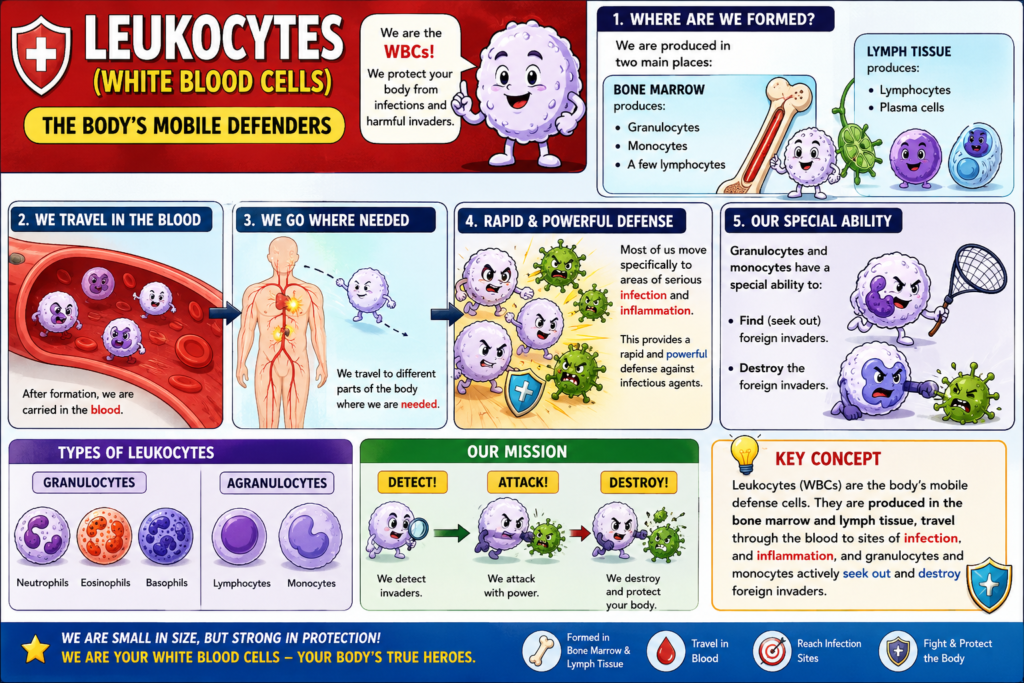
- Leukocytes, also called white blood cells (WBCs), are the mobile protective cells of the body.
- They help protect the body against infection and harmful agents.
- WBCs are formed in two main places:
- Bone marrow produces:
- Granulocytes
- Monocytes
- A few lymphocytes
- Lymph tissue produces:
- Lymphocytes
- Plasma cells
- Bone marrow produces:
- After they are formed, WBCs are carried in the blood.
- They travel to different parts of the body where they are needed.
- The main importance of WBCs is that most of them move specifically to areas of serious infection and inflammation.
- This provides a rapid and powerful defense against infectious agents.
- Granulocytes and monocytes have a special ability to:
- Find (seek out) foreign invaders.
- Destroy the foreign invaders.
Figure
- No figure number is mentioned in the provided text.
Equations
- No mathematical equation is present in the provided text.
Key Concept
Leukocytes (WBCs) are the body’s mobile defense cells. They are produced in the bone marrow and lymph tissue, travel through the blood to sites of infection and inflammation, and granulocytes and monocytes actively seek out and destroy foreign invaders.
General Characteristics of Leukocytes
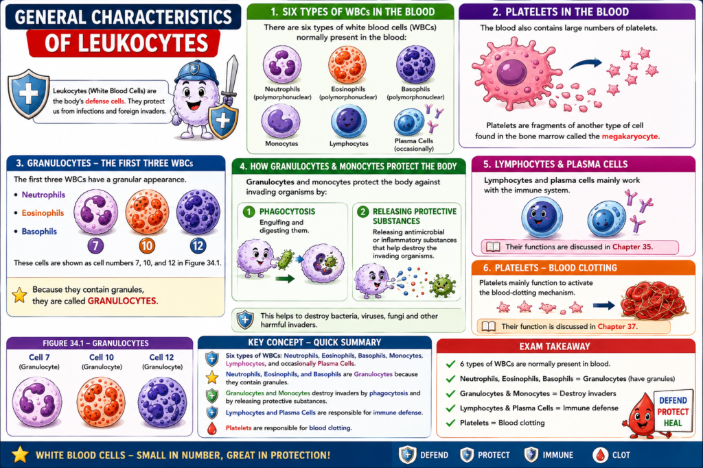
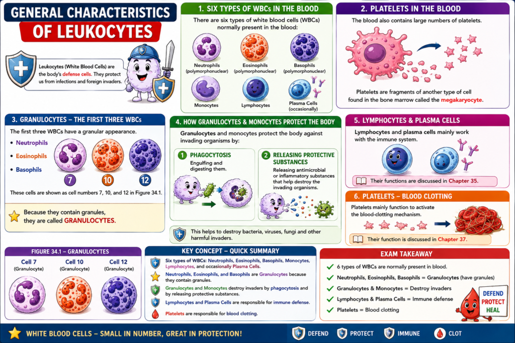
- There are six types of white blood cells (WBCs) normally present in the blood:
- Neutrophils (polymorphonuclear)
- Eosinophils (polymorphonuclear)
- Basophils (polymorphonuclear)
- Monocytes
- Lymphocytes
- Occasionally, plasma cells
- The blood also contains large numbers of platelets.
- Platelets are fragments of another type of cell found in the bone marrow called the megakaryocyte.
- The first three WBCs:
- Neutrophils
- Eosinophils
- Basophils
- These cells have a granular appearance.
- They are shown as cell numbers 7, 10, and 12 in Figure 34.1.
- Because they contain granules, they are called granulocytes.
- Granulocytes and monocytes protect the body against invading organisms by:
- Engulfing and digesting them (phagocytosis).
- Releasing antimicrobial or inflammatory substances that help destroy the invading organisms.
- Lymphocytes and plasma cells mainly work with the immune system.
- Their functions are discussed in Chapter 35.
- Platelets mainly function to activate the blood-clotting mechanism.
- Their function is discussed in Chapter 37.
Figure
- Figure 34.1
- Cell 7 – Granulocyte
- Cell 10 – Granulocyte
- Cell 12 – Granulocyte
Equations
- No mathematical equation is present in the provided text.
Key Concept
There are six types of WBCs in the blood. Neutrophils, eosinophils, and basophils are granulocytes because they contain granules. Granulocytes and monocytes destroy invading organisms by phagocytosis and by releasing protective substances, while lymphocytes and plasma cells are responsible for immune defense, and platelets are responsible for blood clotting.
Concentrations of Different White Blood Cells in Blood
- An adult human normally has about 7,000 white blood cells (WBCs) per microliter of blood.
- In comparison, there are about 5 million red blood cells (RBCs) per microliter of blood.
- The normal percentages of different types of WBCs are approximately:
- Neutrophils: 62.0%
- Eosinophils: 2.3%
- Basophils: 0.4%
- Monocytes: 5.3%
- Lymphocytes: 30.0%
- The normal number of platelets in each microliter of blood is:
- 150,000–450,000 per microliter
- Average: 300,000 per microliter
Key Concept
An adult normally has 7,000 WBCs/µL and 5 million RBCs/µL of blood. Among WBCs, neutrophils are the most abundant (62%), followed by lymphocytes (30%), monocytes (5.3%), eosinophils (2.3%), and basophils (0.4%). The normal platelet count is 150,000–450,000/µL, with an average of 300,000/µL.
Genesis of White Blood Cells
- Early differentiation of the multipotential hematopoietic stem cell into different committed progenitor cells was shown in Figure 33.2.
- Apart from the cells that form red blood cells (RBCs), two major white blood cell (WBC) lineages are formed:
- Myelocytic lineage
- Lymphocytic lineage
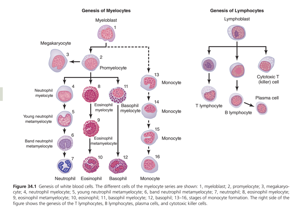
- The left side of Figure 34.1 shows the myelocytic lineage, beginning with the myeloblast.
- The right side of Figure 34.1 shows the lymphocytic lineage, beginning with the lymphoblast.
- Our body is continuously exposed to bacteria, viruses, fungi, and parasites.
- These microorganisms are normally present in the:
- Skin
- Mouth
- Respiratory passages
- Intestinal tract
- Eye lining membranes
- Urinary tract
- Many of these microorganisms can cause serious disease or even death if they enter deeper body tissues.
- The body is also sometimes exposed to other highly infectious bacteria and viruses.
- These can cause severe diseases, such as:
- Pneumonia
- Streptococcal infection
- Typhoid fever
- The body has a special defense system against infectious and toxic agents.
- This defense system consists of:
- Blood leukocytes (white blood cells [WBCs])
- Tissue cells derived from leukocytes
- These cells protect the body in two ways:
- They destroy invading bacteria and viruses by phagocytosis.
- They produce antibodies and sensitized lymphocytes that destroy or inactivate the invading organisms.
- This chapter explains the first defense mechanism (phagocytosis).
- Chapter 35 explains the second defense mechanism (antibodies and sensitized lymphocytes).
- Leukocytes (WBCs) are the mobile protective cells of the body.
- WBCs are formed in two main places:
- Bone marrow forms:
- Granulocytes
- Monocytes
- A few lymphocytes
- Lymph tissue forms:
- Lymphocytes
- Plasma cells
- Bone marrow forms:
- After formation, WBCs are transported in the blood to different parts of the body where they are needed.
- Most WBCs move specifically to areas of serious infection and inflammation.
- This provides a rapid and powerful defense against infectious agents.
- Granulocytes and monocytes have the special ability to seek out and destroy foreign invaders.
- Granulocytes and monocytes are formed only in the bone marrow.
- Lymphocytes and plasma cells are produced mainly in lymphogenous tissues, especially:
- Lymph glands
- Spleen
- Thymus
- Tonsils
- Lymphoid tissue in the bone marrow
- Peyer patches beneath the epithelium of the gut wall
- WBCs formed in the bone marrow are stored in the marrow until they are needed in the circulation.
- When required, various factors cause these WBCs to be released into the blood.
- Normally, the bone marrow stores about three times more WBCs than are circulating in the entire blood.
- This stored amount represents about a 6-day supply of WBCs.
- Most lymphocytes are stored in the lymphoid tissues.
- Only a small number are temporarily transported in the blood.
- Figure 34.1 shows that megakaryocytes (Cell 3) are also formed in the bone marrow.
- Megakaryocytes break into small fragments in the bone marrow.
- These fragments are called platelets (thrombocytes).
- Platelets then enter the blood.
- Platelets are very important for initiating blood clotting.
Figure
- Figure 33.2 – Early differentiation of multipotential hematopoietic stem cells into committed progenitor cells.
- Figure 34.1
- Left side – Myelocytic lineage (begins with myeloblast)
- Right side – Lymphocytic lineage (begins with lymphoblast)
- Cell 3 – Megakaryocyte
Equations
- No mathematical equation is present in the provided text.
Key Concept
White blood cells develop from two major lineages: myelocytic and lymphocytic. Granulocytes and monocytes are formed only in the bone marrow, while lymphocytes and plasma cells are mainly produced in lymphoid tissues. WBCs protect the body by phagocytosis and by forming antibodies and sensitized lymphocytes. Bone marrow stores about three times the circulating WBCs, providing approximately a 6-day reserve, and megakaryocytes produce platelets, which initiate blood clotting.
Genesis of White Blood Cells (WBCs) – Easiest Concept (Guyton Physiology Figure 34.1)
One Main Idea to Remember
All white blood cells (WBCs) are produced in the bone marrow from stem cells.
After that, they divide into 2 major pathways (families):
Stem Cell
      │
      ├──────────────► Myelocytic Lineage (Bone Marrow Cells)
      │
      └──────────────► Lymphocytic Lineage (Immune Cells)
1. Myelocytic Lineage (Left Side)
Starts with:
Myeloblast (Cell 1)
⬇️
Promyelocyte (Cell 2)
This is the common parent cell that gives rise to almost all granulocytes and monocytes.
From here, 4 different cells are formed.
A. Neutrophil Formation
This is the longest maturation process.
Myeloblast
      ↓
Promyelocyte
      ↓
Neutrophil Myelocyte
      ↓
Young Metamyelocyte
      ↓
Band Neutrophil
      ↓
Mature Neutrophil
Easy Trick
My → Pro → Myelo → Young → Band → Mature
Function
✅ First responder to bacterial infection
✅ Eats bacteria (Phagocytosis)
✅ Most abundant WBC (60–70%)
B. Eosinophil Formation
Promyelocyte
      ↓
Eosinophil Myelocyte
      ↓
Eosinophil Metamyelocyte
      ↓
Mature Eosinophil
Function
✅ Fights parasites
✅ Controls allergic reactions
C. Basophil Formation
Promyelocyte
      ↓
Basophil Myelocyte
      ↓
Mature Basophil
Function
Releases
- Histamine
- Heparin
Responsible for
- Allergy
- Inflammation
D. Monocyte Formation
Myeloblast
      ↓
Promyelocyte
      ↓
Several Monocyte Stages
      ↓
Mature Monocyte
Function
Largest WBC
Leaves blood and becomes
➡️ Macrophage
Macrophages eat
- Bacteria
- Dead cells
- Foreign material
E. Megakaryocyte Formation
The figure also shows
Promyelocyte
      ↓
Megakaryocyte
Function
Megakaryocyte breaks into
➡️ Platelets
Platelets help in
✔ Blood clotting
Summary of Myelocytic Lineage
| Cell Produced | Main Function |
|---|---|
| Neutrophil | Bacteria killer |
| Eosinophil | Parasites & allergy |
| Basophil | Histamine & allergy |
| Monocyte | Becomes macrophage |
| Megakaryocyte | Produces platelets |
2. Lymphocytic Lineage (Right Side)
Starts from
LymphoblastIt produces three important immune cells.
A. T Lymphocyte
Lymphoblast
      ↓
T Lymphocyte
Function
Cell-mediated immunity
Kills infected cells
Helps fight viruses
B. Cytotoxic (Killer) T Cell
Lymphoblast
      ↓
Cytotoxic T Cell
Function
Directly destroys
- Virus-infected cells
- Cancer cells
- Transplanted foreign cells
C. B Lymphocyte
Lymphoblast
      ↓
B Lymphocyte
      ↓
Plasma Cell
Plasma Cell Function
Produces
🛡️ Antibodies (Immunoglobulins)
This is called
Humoral Immunity
Easy Difference
T Cells
Fight using cells
➡️ Attack infected cells directly
B Cells
Fight using antibodies
➡️ Plasma cells make antibodies
Whole Figure in One Flow
Stem Cell
       │
 ┌─────┴──────────┐
 │                │
Myeloblast    Lymphoblast
 │                │
 │                ├────────► T Lymphocyte
 │                │
 │                ├────────► Killer T Cell
 │                │
 │                └────────► B Lymphocyte
 │                              │
 │                              ▼
 │                         Plasma Cell
 │
 ├────────► Neutrophil
 │
 ├────────► Eosinophil
 │
 ├────────► Basophil
 │
 ├────────► Monocyte → Macrophage
 │
 └────────► Megakaryocyte → Platelets
Super Easy Memory Trick
MYELO = “Many Body Defenders”
Produces:
- N = Neutrophil → Bacteria
- E = Eosinophil → Parasites
- B = Basophil → Allergy
- M = Monocyte → Macrophage
- P = Platelets (via Megakaryocyte)
Mnemonic: “Never Eat Bad Meat Please”
- N = Neutrophil
- E = Eosinophil
- B = Basophil
- M = Monocyte
- P = Platelets
LYMPHO = “Specific Immunity”
Produces:
- T Cell → Cell-mediated immunity
- Killer T Cell → Kills infected/cancer cells
- B Cell → Makes Plasma Cells
- Plasma Cell → Produces antibodies
Exam Points (Most Important)
- All WBCs originate from hematopoietic stem cells in the bone marrow.
- Two major lineages: Myelocytic and Lymphocytic.
- Myeloblast gives rise to neutrophils, eosinophils, basophils, monocytes, and megakaryocytes.
- Lymphoblast gives rise to T lymphocytes, B lymphocytes, cytotoxic T cells, and plasma cells.
- Neutrophils are the first responders against bacterial infections.
- Eosinophils fight parasites and participate in allergic reactions.
- Basophils release histamine and heparin.
- Monocytes leave the bloodstream and become macrophages in tissues.
- B lymphocytes differentiate into plasma cells, which produce antibodies.
- T lymphocytes provide cell-mediated immunity, while cytotoxic T cells directly kill infected and abnormal cells.
Life Span of White Blood Cells
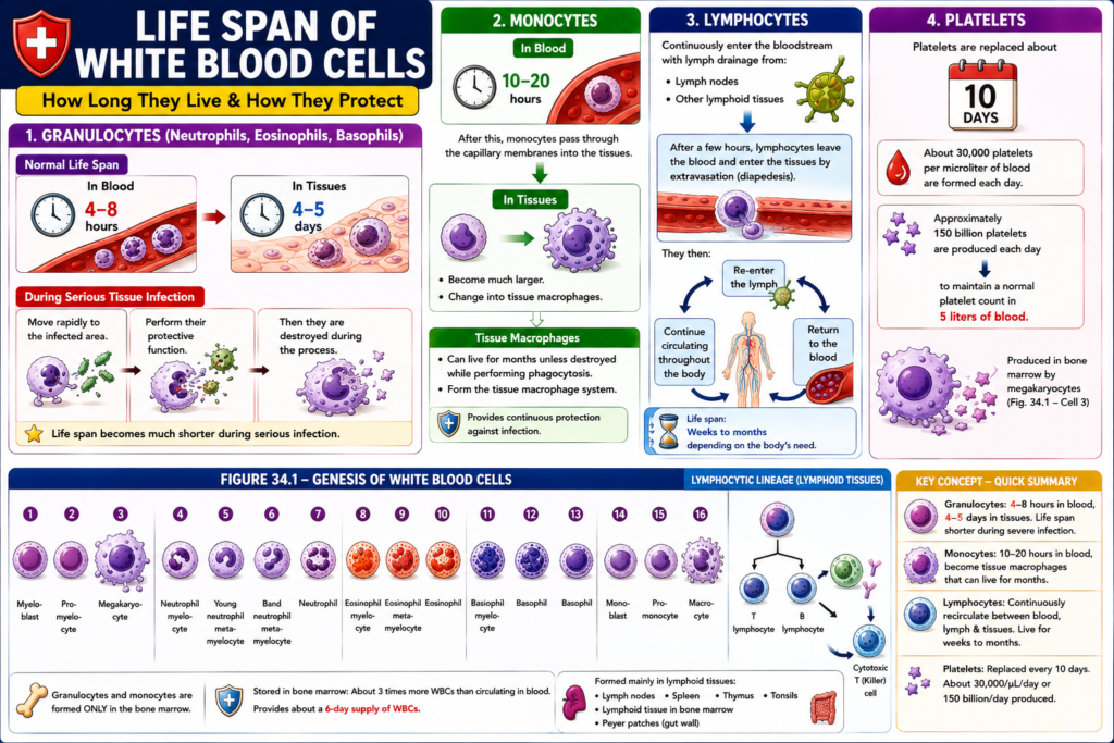
- After being released from the bone marrow, granulocytes normally:
- Circulate in the blood for 4–8 hours.
- Live in the tissues for another 4–5 days where they are needed.
- During serious tissue infection:
- The life span of granulocytes becomes much shorter.
- They move rapidly to the infected area.
- They perform their protective function.
- They are then destroyed during the process.
- Monocytes remain in the blood for 10–20 hours.
- After this, monocytes pass through the capillary membranes into the tissues.
- In the tissues, monocytes:
- Become much larger.
- Change into tissue macrophages.
- Tissue macrophages can live for months unless they are destroyed while performing phagocytosis.
- Tissue macrophages form the tissue macrophage system.
- This system provides continuous protection against infection.
- Lymphocytes continuously enter the bloodstream with lymph drainage from:
- Lymph nodes
- Other lymphoid tissues
- After a few hours, lymphocytes leave the blood and enter the tissues by extravasation (diapedesis).
- They then:
- Re-enter the lymph.
- Return to the blood.
- Continue circulating throughout the body.
- The life span of lymphocytes is weeks to months, depending on the body’s need.
- Platelets are replaced about once every 10 days.
- About 30,000 platelets per microliter of blood are formed each day.
- Approximately 150 billion platelets are produced each day to maintain a normal platelet count in 5 liters of blood.
Figure
- Figure 34.1 – Genesis of White Blood Cells
- 1 – Myeloblast
- 2 – Promyelocyte
- 3 – Megakaryocyte
- 4 – Neutrophil myelocyte
- 5 – Young neutrophil metamyelocyte
- 6 – Band neutrophil metamyelocyte
- 7 – Neutrophil
- 8 – Eosinophil myelocyte
- 9 – Eosinophil metamyelocyte
- 10 – Eosinophil
- 11 – Basophil myelocyte
- 12 – Basophil
- 13–16 – Stages of monocyte formation
- Right side:
- Lymphoblast
- T lymphocyte
- B lymphocyte
- Plasma cell
- Cytotoxic T (killer) cell
Equations
- No mathematical equation is present in the provided text.
Key Concept
- Granulocytes: 4–8 hours in blood + 4–5 days in tissues (shorter during severe infection).
- Monocytes: 10–20 hours in blood, then become tissue macrophages that can live for months.
- Lymphocytes: Continuously recirculate between blood, tissues, and lymph and live for weeks to months.
- Platelets: Replaced every 10 days, with about 30,000/µL/day or 150 billion/day produced to maintain a normal platelet count.
MADE BY EASIEST SELF LEARNING CEO AND FOUNDER DR SHEEN
